• Home
  • About
  • Articles
  • Books
  • Contact

Veterinary Radiology

Teaching and learning about veterinary diagnostic imaging.

  • Modality
    • Radiographs
    • Ultrasound
    • CT/MRI
  • Region
    • Thorax
    • Abdomen
    • Musculoskeletal
    • Neurologic
  • Species
    • Canine
    • Feline
    • Equine
    • Exotic

8 year old German Shepherd cross

March 30, 2015 By Allison Zwingenberger

This week’s case is an 8 year old male neutered German Shepherd cross with a 48-hour history of lethargy, pale mucous membranes, and a distended abdomen. Bloodwork indicates anemia, neutrophilia, thrombocytopenia, increased ALT and AST, increased CK and GGT, decreased cholesterol and T4.

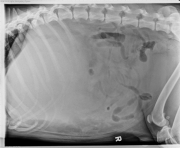
R LAT Abdomen
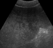
Ultrasound 1
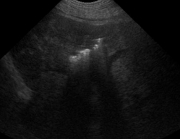
Ultrasound 2

Show findings...

Findings

There is increased soft tissue opacity in the cranial abdomen with caudal displacement of the stomach and small intestine. The abdomen is distended, and there is mottled peritoneal detail in the cranioventral region. There is also a gas lucency overlying the dorsal portion of the liver. This may be overlying bowel, or be within the hepatic parenchyma.

The ultrasound exam shows a liver infiltrated with multiple masses. The masses are of mixed echogenicity, and deform the capsule of the liver. Within one of the masses, there are multiple hyperechoic foci with distal “dirty” acoustic shadowing. The dirty shadowing indicates gas, while clean shadowing would indicate mineralization.

Show differential diagnosis…

Differential Diagnosis

  • Hepatic mass
  • Splenic mass

Show diagnosis…

Diagnosis

Hepatic carcinoma with suppurative effusion and hemorrhage.

Show discussion…

Discussion

The cranial location of the mass effect makes the most likely differential diagnosis a hepatic mass. A splenic mass is possible, however these are more often in the mid abdomen and better delineated. The gastric axis is faintly visible caudal to the mass, supporting a hepatic origin.

The distended abdomen and poor detail are diagnostic of peritoneal effusion. The compression of abdominal organs is also contributing to poor viewing of serosal surfaces.

Hepatic abscesses in dogs can be caused by liver lobe torsion, infection secondary to diabetes mellitus, systemic sepsis, or within masses. The infection is often anerobic, with E. coli spreading from the gastrointestinal tract.

Show references…

References

  1. Sato AF, Solano M. Radiographic diagnosis: liver lobe entrapment and associated emphysematous hepatitis. Veterinary Radiology & Ultrasound 1998;39:123-124.
  2. Lord PF, Carb A, Halliwell WH, et al. Emphysematous hepatic abscess associated with trauma, necrotic hepatic nodular hyperplasia and adenoma in a dog: a case history report. Veterinary Radiology 1982;23:46-49.
  3. Grooters AM, Sherding RG, Biller DS, et al. Hepatic abscesses associated with diabetes mellitus in two dogs. J Vet Intern Med 1994;8:203-206.
  4. Grooters AM, Sherding RG, Johnson SE. Hepatic abscesses in dogs. Compendium on Continuing Education for the Practicing Veterinarian 1995;17:833-836, 838, 840.
  5. Downs MO, Miller MA, Cross AR, et al. Liver lobe torsion and liver abscess in a dog. J Am Vet Med Assoc 1998;212:678-680.
  6. Farrar ET, Washabau RJ, Saunders HM. Hepatic abscesses in dogs: 14 cases (1982-1994). J Am Vet Med Assoc 1996;208:243-247.

 
Case originally posted on January 29, 2007
Filed Under: Abdomen, Canine, Case of the Day, Radiographs, Ultrasound

  • Bloglovin
  • Facebook
  • RSS
  • Twitter
AtlasCover787x1024 amazon-availablenow

Veterinary Radiology News

Sign up for alerts about new cases and newsletters.

We will respect your privacy.

Archives

Recent Comments

Tags

abscess adenocarcinoma bronchiectasis carcinoma cardiomyopathy coccidioidomycosis discospondylitis elbow dysplasia eosinophilic bronchopneumopathy erosive polyarthritis feline infectious peritonitis FIP foreign body fracture gastric dilation gastric foreign body gastrointestinal GDV heart failure hemangiosarcoma hiatal hernia histiocytic sarcoma hypertrophic cardiomyopathy intestinal foreign body linear foreign body lymphoma megaesophagus osteochondrosis osteomyelitis osteosarcoma patent ductus arteriosus PDA pericardial effusion pneumonia pneumothorax polyarthritis PPDH pulmonary pulmonary abscess pulmonary adenocarcinoma pulmonary carcinoma sarcoma thymoma tracheal collapse vascular ring anomaly

Copyright © 2025 · Metro Pro Theme On Genesis Framework · WordPress · Log in