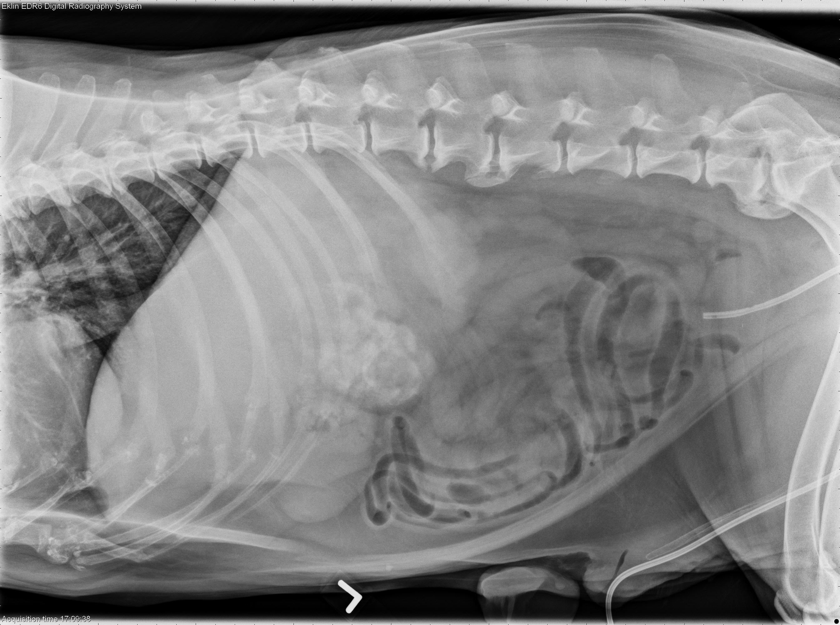
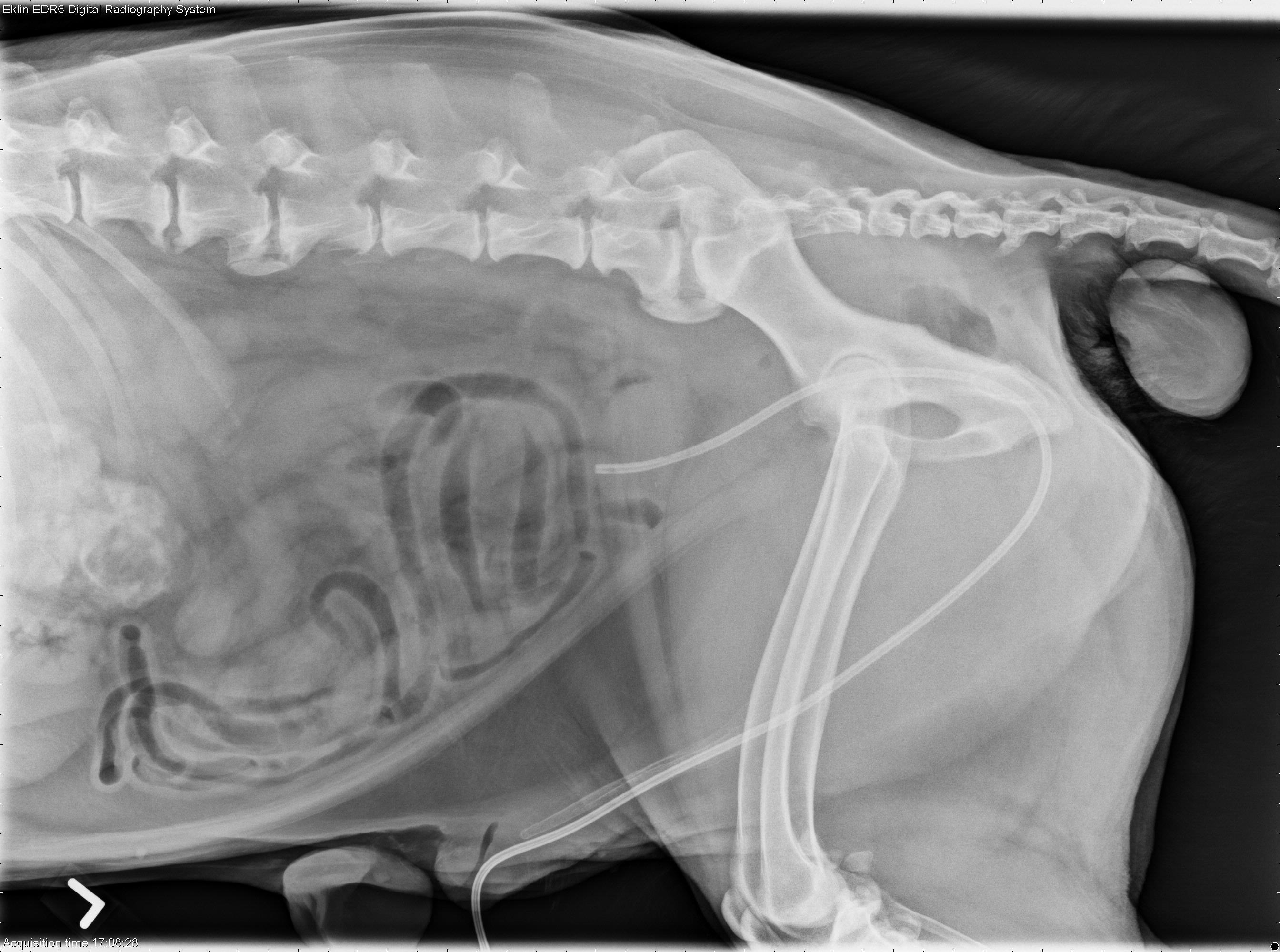
Today’s case is a 13-year-old male neutered German Shepherd that has been losing weight for three months, and has had a 7-day history of vomiting, diarrhea, lethargy, inappetence, decreased drinking, and straining to defecate and urinate. What are your findings?
Findings
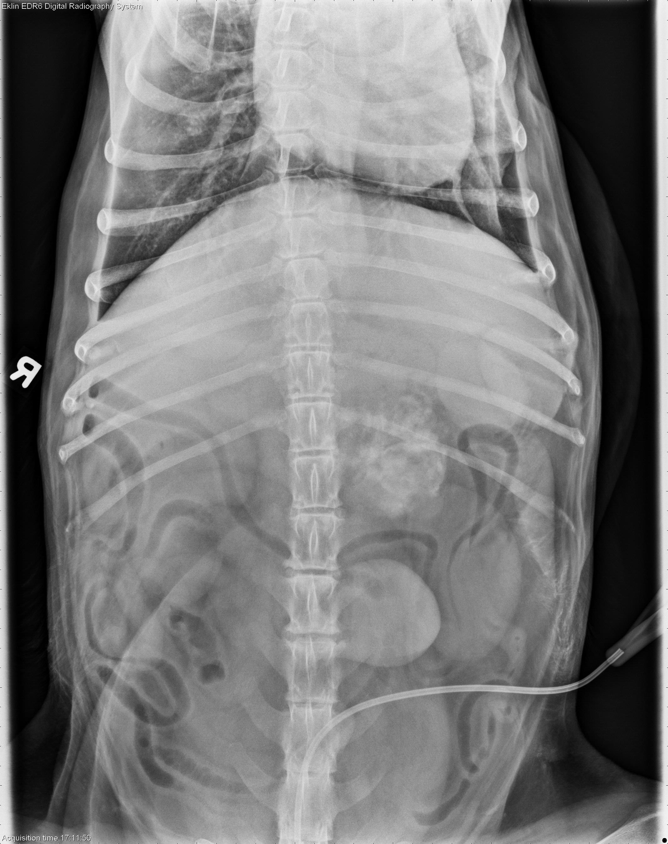
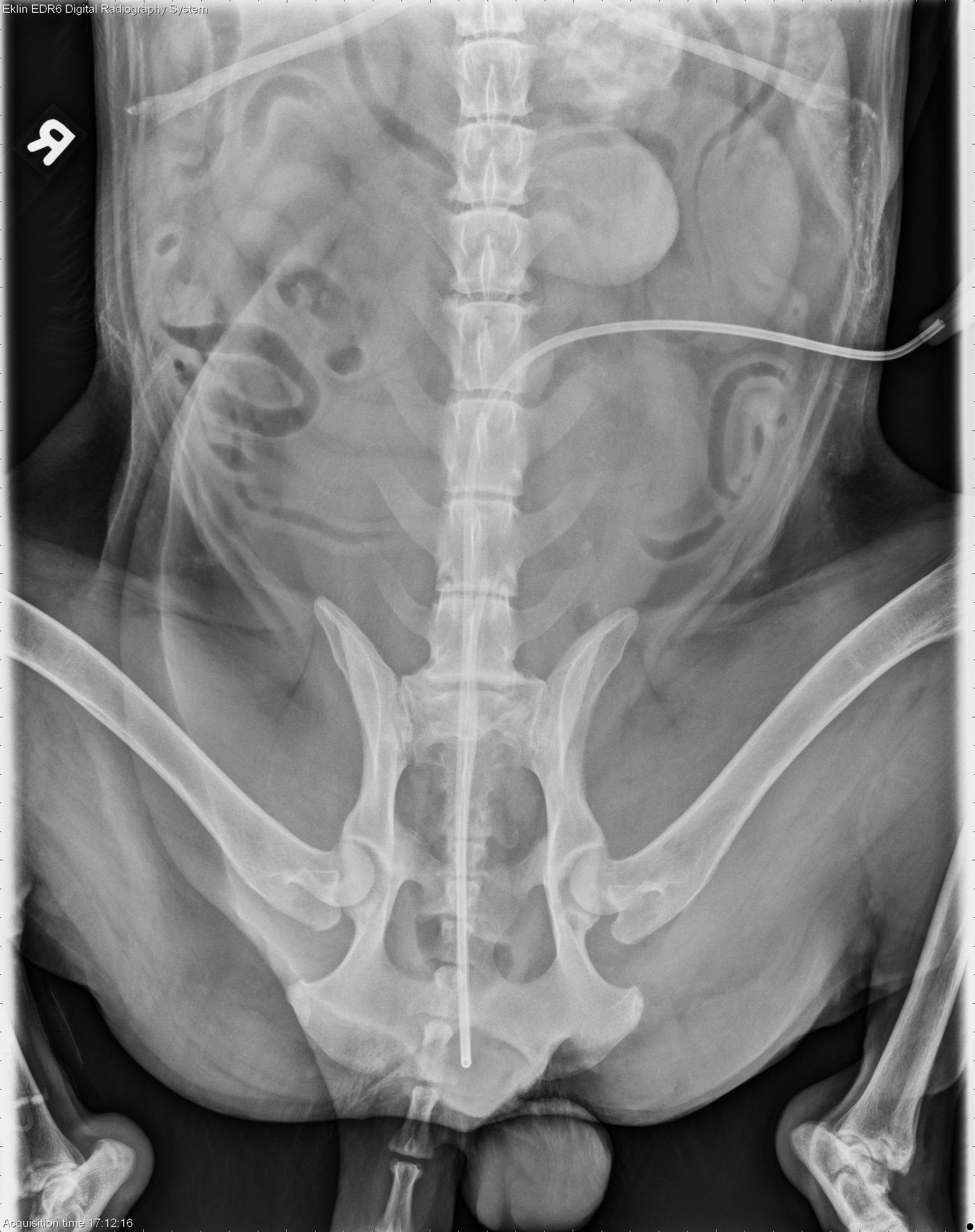
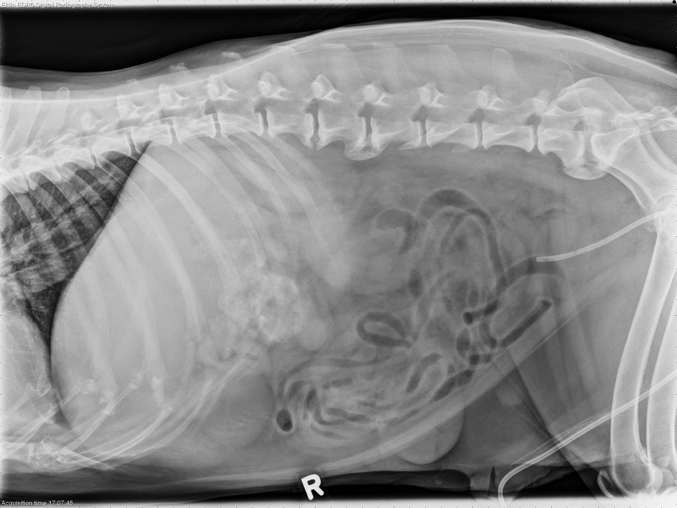
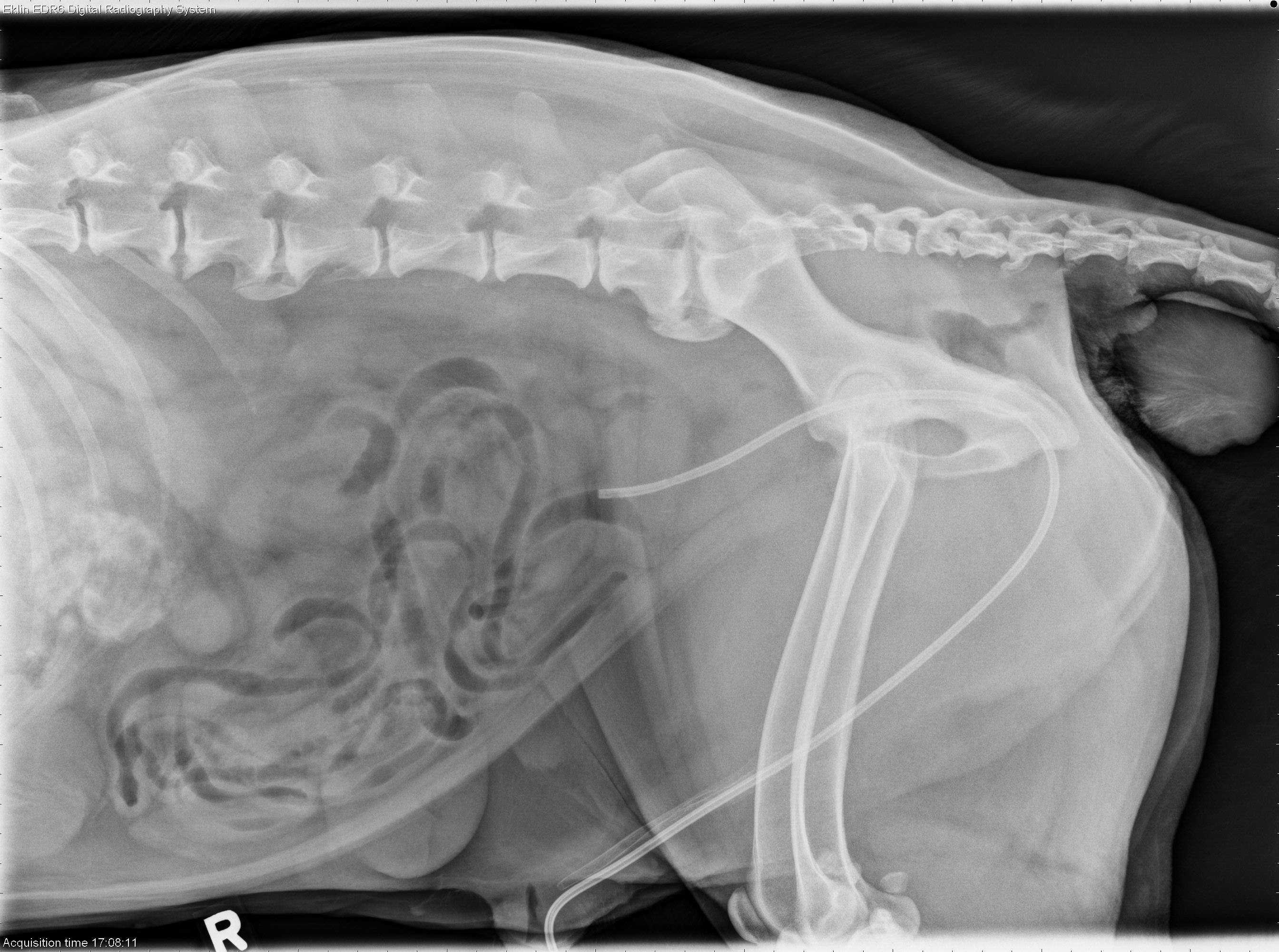
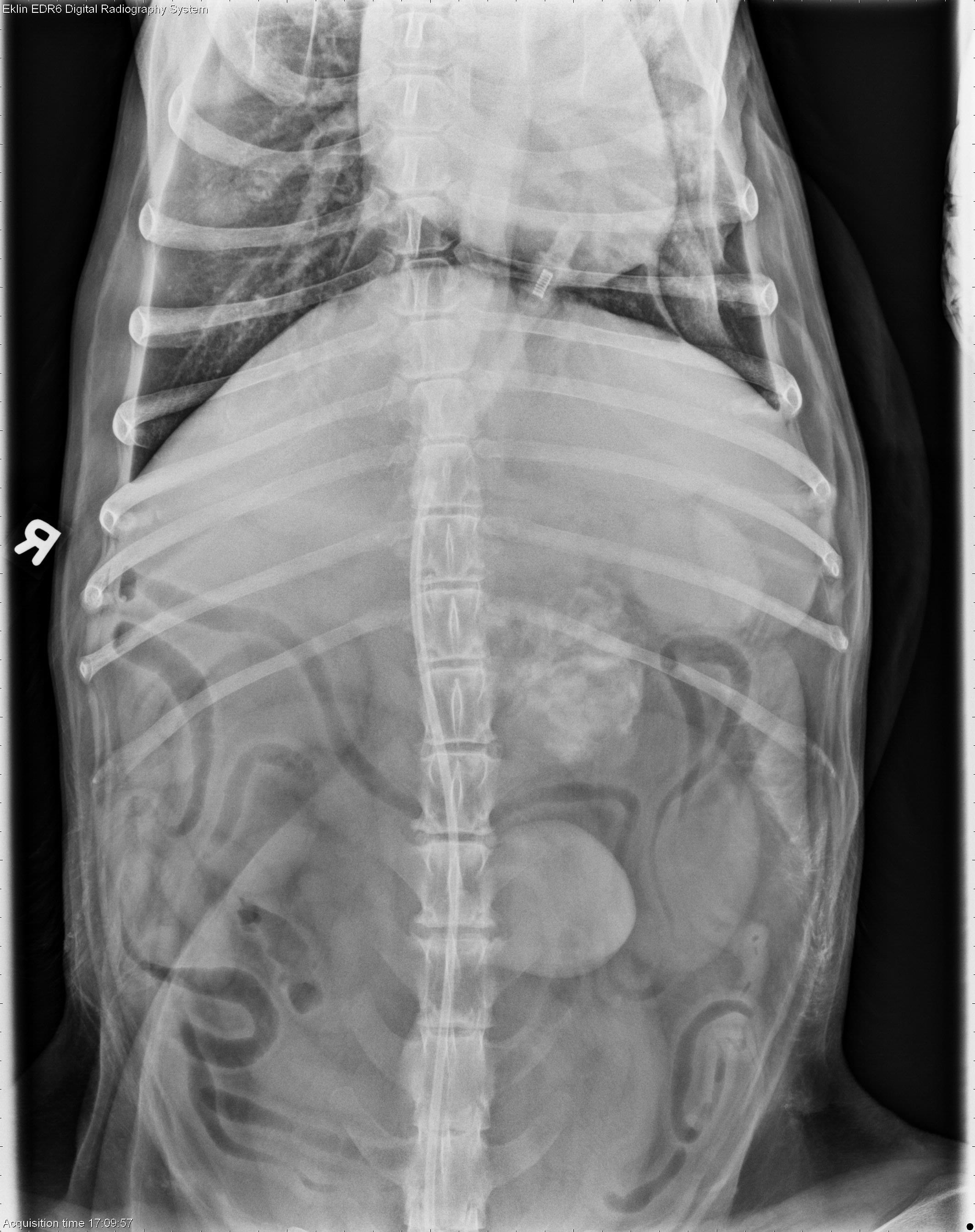
There is an ill-defined, rounded region of heterogeneous mineral and soft tissue opacity within the cranial abdomen near midline. The mass is positioned to the left of midline, cranial to the left kidney on the v/d projection, and dorsal to the pylorus on both the left and right lateral projections. The liver appears enlarged with ill-defined borders on all views.
The kidneys are mildly enlarged. A urinary catheter is present which terminates at the apex of the urinary bladder. There are large, rounded soft tissue masses on the perineum and prepuce. There is mild multifocal spondylosis deformans which is worse at the L-S space.
Differential Diagnosis
- liver mass (most likely primary neoplasia, less likely granuloma)
- renal enlargement (hydronephrosis, pyelonephritis, neoplasia)
Diagnosis
On abdominal ultrasound, a large mineralized liver mass was identified. There was also a mass in the urinary bladder causing bilateral ureteral obstruction and hydronephrosis. The patient was euthanized at the owner’s request, without tissue sampling.