• Home
  • About
  • Articles
  • Books
  • Contact

Veterinary Radiology

Teaching and learning about veterinary diagnostic imaging.

  • Modality
    • Radiographs
    • Ultrasound
    • CT/MRI
  • Region
    • Thorax
    • Abdomen
    • Musculoskeletal
    • Neurologic
  • Species
    • Canine
    • Feline
    • Equine
    • Exotic

Skin mass or pulmonary nodule?

September 4, 2007 By Allison Zwingenberger

A dog with multiple small skin masses on the thoracic body wall comes in for a metastasis check. How do you decide whether the soft tissue opacities you can see are on the surface or in the lung parenchyma? There are a few ways you can try to distinguish them.

Sharpness of the margin

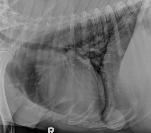
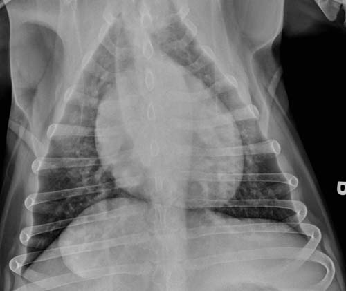
We can see pulmonary nodules or skin masses as separate from lung or skin because they are surrounded by air, giving a soft-tissue/gas interface. Nodules in the lung are also surrounded by small vessels and pulmonary tissue, as well as the rest of the thorax. This means that skin masses that are surrounded by air only have much sharper margination than lung nodules. Compare the sharpness of the body wall mass on the caudoventral left thorax to the pulmonary nodules in the images. This dog has metastases from the large liver mass visible in the cranial abdomen as well as a lipoma.

Completeness of the margin

Because body wall masses are attached to the skin, the visible margin is usually incomplete. The air surrounds the mass on two or three sides, but not circumferentially. Lung nodules have margins that are visible for 360 degrees unless they are silhouetting with another structure. Check multiple nodules for completeness of the margins.

Use a marker

If you still can’t tell if the soft tissue structure you are looking at is a skin mass or a pulmonary nodule, palpate the dog and place a marker on the skin mass. A dab of liquid barium or metallic adhesive markers both work well.

Skin mass or pulmonary nodule?Skin mass or pulmonary nodule?

Filed Under: Uncategorized

  • Bloglovin
  • Facebook
  • RSS
  • Twitter
AtlasCover787x1024 amazon-availablenow

Veterinary Radiology News

Sign up for alerts about new cases and newsletters.

We will respect your privacy.

Archives

Recent Comments

Tags

abscess adenocarcinoma bronchiectasis carcinoma cardiomyopathy coccidioidomycosis discospondylitis elbow dysplasia eosinophilic bronchopneumopathy erosive polyarthritis feline infectious peritonitis FIP foreign body fracture gastric dilation gastric foreign body gastrointestinal GDV heart failure hemangiosarcoma hiatal hernia histiocytic sarcoma hypertrophic cardiomyopathy intestinal foreign body linear foreign body lymphoma megaesophagus osteochondrosis osteomyelitis osteosarcoma patent ductus arteriosus PDA pericardial effusion pneumonia pneumothorax polyarthritis PPDH pulmonary pulmonary abscess pulmonary adenocarcinoma pulmonary carcinoma sarcoma thymoma tracheal collapse vascular ring anomaly

Copyright © 2025 · Metro Pro Theme On Genesis Framework · WordPress · Log in