We all know that there are artifacts on just about every ultrasound image we look at. The physics of the sound waves interacting with tissues is complicated! But many of these artifacts need to be recognized and acknowledged so that you can move on with interpreting the images. The mirror image artifact is one that you’ll see in the cranial part of the abdomen.
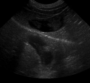 Ultrasound images are formed by the transducer producing the beam, and also detecting that beam as it is reflected from tissue interfaces. The transducer calculates how much time has passed since that particular pulse left, and uses the time to place the image at the correct depth on the screen. It always assumes that the ultrasound pulse traveled in a straight line.
Highly reflective, curved surfaces, like the diaphragm, can reflect that pulse of sound in a different direction before it returns to the transducer. So the transducer places that portion of the image deeper than it actually is in the body. In the case of the diaphragm-lung interface, the image gets mapped to the far side of the diaphragm where the lungs are. It can look like there is a diaphragmatic hernia, with liver present in the thorax, or like there is consolidation of a lung lobe. In this example, the liver parenchyma and gallbladder are mirrored into the thorax with the sharp hyperechoic line of the diaphragm separating them.
Recent Comments