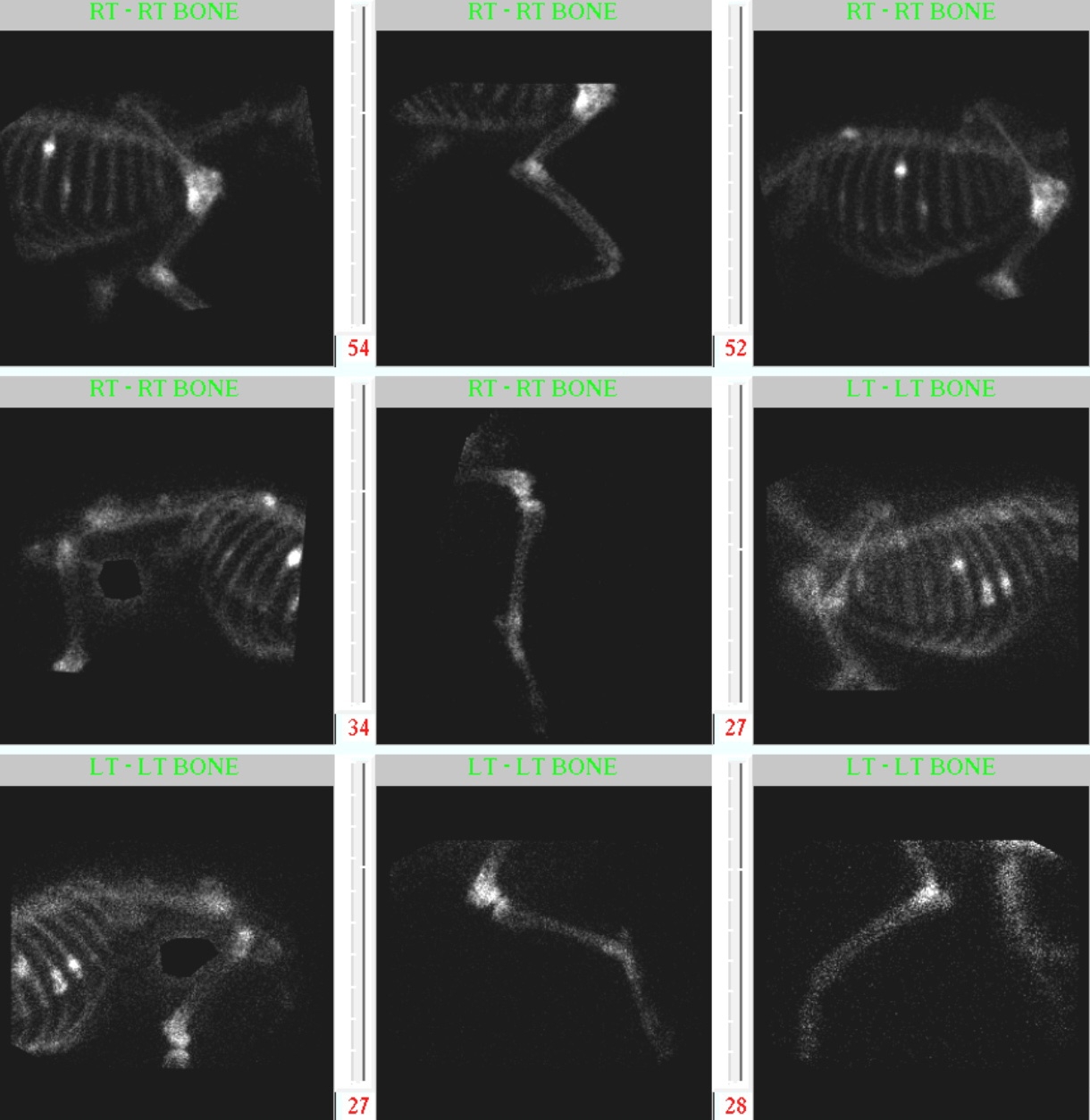
This is a 6-year-old female neutered Rottweiler with right forelimb lameness for one month. Post your interpretations in the comments section!
Findings
The cardiovascular and pulmonary structures appear to be within normal limits. The proximal humerus of the right shoulder is lytic with a smoothly margined area of new bone seen extending from the neck of the humeral head to the proximal diaphysis along the caudal margin. The proximal right 7th, distal right 6th, distal left 8th and distal left 9th ribs also appear to be affected with focal areas of expansile, mixed opacity new bone and lysis. Multiple circular lytic areas without surrounding sclerosis are seen scattered throughout both humerii and the thoracic spinous processes. A transverse fracture with a centralized lucent area and surrounding bony remodeling is seen in the dorsal third of the third spinous process. No pulmonary nodules are noted.
Differential Diagnoses
The appearance of the proximal right humerus is consistent with a primary bone tumor. The polyostotic lytic lesions are most likely metastasis from the primary bone tumor.  Suspect pathologic fracture of the third thoracic spinous process.
Nuclear Scintigraphy
A Â survey musculoskeletal bone scan was performed to confirm the sites of metastatic disease. Several sites are present in the left and right ribs. The 10th thoracic vertebra also has increased radiopharmaceutical uptake with lesser uptake in the lumbar third vertebrae. The multiple small humeral lesions are not apparent on the images however the primary humeral lesion has strong uptake.
Diagnosis
Primary osteosarcoma of the humerus with metastatic spread to the axial and appendicular skeleton.

Recent Comments