Today’s case is a 12 year old male neutered Border Collie who underwent routine thoracic radiographs.
Findings
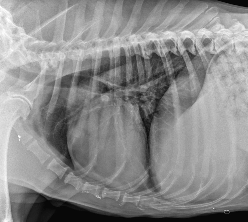
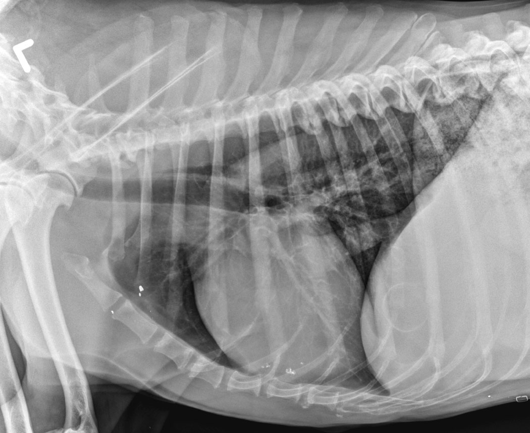
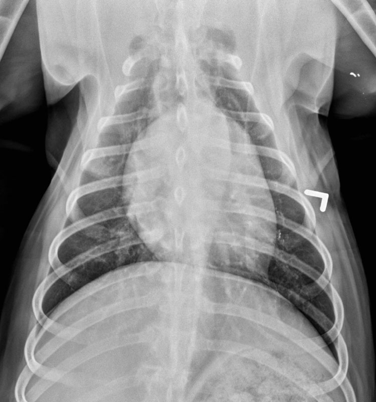
There are multiple thin-walled, air filled lucencies throughout the lungs. These bullae are visible on all projections. They are best seen superimposed over the diaphragm.
Differential Diagnosis
- bulla (emphysema, congenital)
- pulmonary cyst
- bronchogenic cyst
- parasitic (Paragonimus kellicotti)
- abscess
- pneumatocele (secondary to pneumonia or trauma)
- neoplasia
Diagnosis
These lesions are most consistent with congenital bullae or bronchogenic cysts.
Discussion
Pulmonary bullae are occasional incidental findings on thoracic radiographs. They are thin-walled and often multiple, with distribution in many lung lobes. Thinner-walled lesions would be more compatible with emphysema, and thick or irregularly walled lesions are more likely to be infectious, neoplastic or parasitic in origin. Any bullous lesion can rupture and cause pneumothorax.