• Home
  • About
  • Articles
  • Books
  • Contact

Veterinary Radiology

Teaching and learning about veterinary diagnostic imaging.

  • Modality
    • Radiographs
    • Ultrasound
    • CT/MRI
  • Region
    • Thorax
    • Abdomen
    • Musculoskeletal
    • Neurologic
  • Species
    • Canine
    • Feline
    • Equine
    • Exotic

12 year old Pit Bull Terrier

February 15, 2016 By Allison Zwingenberger

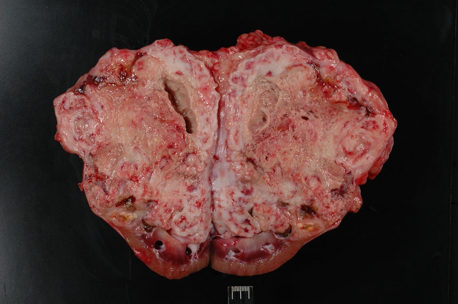
Today’s case is a 12 year old MC Pit Bull Terrier with tetraparesis and ataxia. What are your findings?

R LAT Abdomen
VD Abdomen

Show findings...

Findings

There is focal poor detail in the dorsal and left mid-abdomen. The poor detail is caused by a soft tissue mass with areas of amorphic mineralization. The mass occupies the area of the left kidney, and no normal left kidney is visible. The mass represents a focal enlargement of the left kidney. The colon is displaced medially on the v/d projection. The liver is moderately enlarged.

Show differential diagnosis…

Differential Diagnosis

  • Primary neoplasia
  • Metastatic neoplasia
  • Granuloma

Show diagnosis…

Diagnosis

Concurrent primary renal carcinoma and osteosarcoma (confirmed by immunohistochemistry)

Show discussion…

Discussion

This lesion has all the features of a renal mass, with location and displacement of neighboring organs, and the focal enlargement indicates a mass lesion. The final diagnosis is unusual with two apparent primary renal tumors. The osteosarcoma was responsible for the mineralization of the mass. No reason for the ataxia and paresis was found at post mortem.

cod160215-3

Filed Under: Abdomen, Canine, Case of the Day, Radiographs

  • Bloglovin
  • Facebook
  • RSS
  • Twitter
AtlasCover787x1024 amazon-availablenow

Veterinary Radiology News

Sign up for alerts about new cases and newsletters.

We will respect your privacy.

Archives

Recent Comments

Tags

abscess adenocarcinoma bronchiectasis carcinoma cardiomyopathy coccidioidomycosis discospondylitis elbow dysplasia eosinophilic bronchopneumopathy erosive polyarthritis feline infectious peritonitis FIP foreign body fracture gastric dilation gastric foreign body gastrointestinal GDV heart failure hemangiosarcoma hiatal hernia histiocytic sarcoma hypertrophic cardiomyopathy intestinal foreign body linear foreign body lymphoma megaesophagus osteochondrosis osteomyelitis osteosarcoma patent ductus arteriosus PDA pericardial effusion pneumonia pneumothorax polyarthritis PPDH pulmonary pulmonary abscess pulmonary adenocarcinoma pulmonary carcinoma sarcoma thymoma tracheal collapse vascular ring anomaly

Copyright © 2025 · Metro Pro Theme On Genesis Framework · WordPress · Log in