• Home
  • About
  • Articles
  • Books
  • Contact

Veterinary Radiology

Teaching and learning about veterinary diagnostic imaging.

  • Modality
    • Radiographs
    • Ultrasound
    • CT/MRI
  • Region
    • Thorax
    • Abdomen
    • Musculoskeletal
    • Neurologic
  • Species
    • Canine
    • Feline
    • Equine
    • Exotic

14 year old Labrador

September 14, 2015 By Allison Zwingenberger

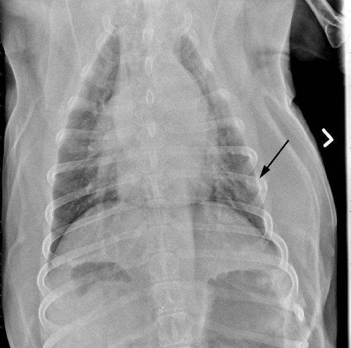
Today’s case is a 14 year old male neutered Labrador Retriever with a two day history of hematuria with clots. Post your interpretations in the comments!

R LAT Thorax
R LAT Thorax
VD Thorax
Ultrasound 1
Ultrasound 2
Ultrasound 3

Show findings...

Findings

Ultrasound:

There is a large, irregularly bordered, broad-based, mineralized mass at the apex of the urinary bladder. There is loss of wall layering adjacent to this mass. There is blood flow detected within this mass. The trigone is within normal limits.

Radiographs:

There are moderate diffuse bronchointerstitial infiltrates throughout the lungs, relatively similar to the previous exam. The cardiovascular structures again appear within normal limits. There is lysis with adjacent soft tissue opacity of the left 7th rib. In the viewable abdomen, the stomach is moderately gas distended. There are multiple hemoclips present ventrally.

Show differential diagnosis…

Differential Diagnosis

Ultrasound:

Large mineralized mass at the apex of the urinary bladder. Differentials include a neoplasia such as a transitional cell carcinoma or severe polypoid cystitis.

Radiographs:

Moderate diffuse bronchointerstitial infiltrates, likely secondary to obesity and age related changes. Lytic lesion of the left 7th rib which likely represents a metastatic lesion.

VD Thorax-annotated

Show diagnosis…

Diagnosis

Transitional cell carcinoma with rib metastasis

Case originally posted on July 23, 2009

Filed Under: Abdomen, Canine, Case of the Day, Radiographs, Ultrasound

Comments

  1. emilycarlo says

    September 14, 2015 at 7:33 am

    Hello to all. I would like to check with the colleagues about a discrete alveolar pattern, since mainly on VD projection, some air bronchograms can be seen.

  • Bloglovin
  • Facebook
  • RSS
  • Twitter
AtlasCover787x1024 amazon-availablenow

Veterinary Radiology News

Sign up for alerts about new cases and newsletters.

We will respect your privacy.

Archives

Recent Comments

Tags

abscess adenocarcinoma bronchiectasis carcinoma cardiomyopathy coccidioidomycosis discospondylitis elbow dysplasia eosinophilic bronchopneumopathy erosive polyarthritis feline infectious peritonitis FIP foreign body fracture gastric dilation gastric foreign body gastrointestinal GDV heart failure hemangiosarcoma hiatal hernia histiocytic sarcoma hypertrophic cardiomyopathy intestinal foreign body linear foreign body lymphoma megaesophagus osteochondrosis osteomyelitis osteosarcoma patent ductus arteriosus PDA pericardial effusion pneumonia pneumothorax polyarthritis PPDH pulmonary pulmonary abscess pulmonary adenocarcinoma pulmonary carcinoma sarcoma thymoma tracheal collapse vascular ring anomaly

Copyright © 2025 · Metro Pro Theme On Genesis Framework · WordPress · Log in