• Home
  • About
  • Articles
  • Books
  • Contact

Veterinary Radiology

Teaching and learning about veterinary diagnostic imaging.

  • Modality
    • Radiographs
    • Ultrasound
    • CT/MRI
  • Region
    • Thorax
    • Abdomen
    • Musculoskeletal
    • Neurologic
  • Species
    • Canine
    • Feline
    • Equine
    • Exotic

1 year old Pit Bull Terrier

March 16, 2015 By Allison Zwingenberger

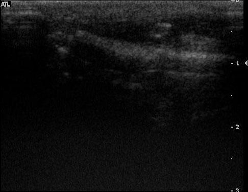
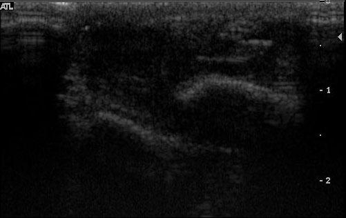
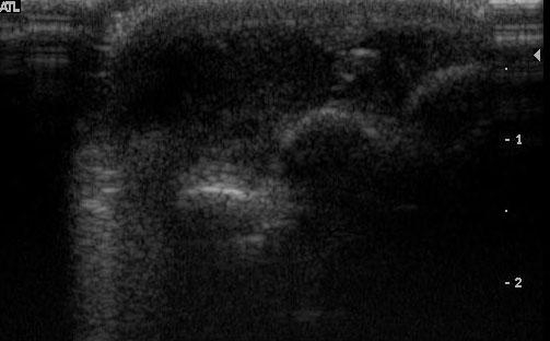
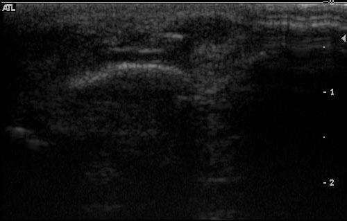
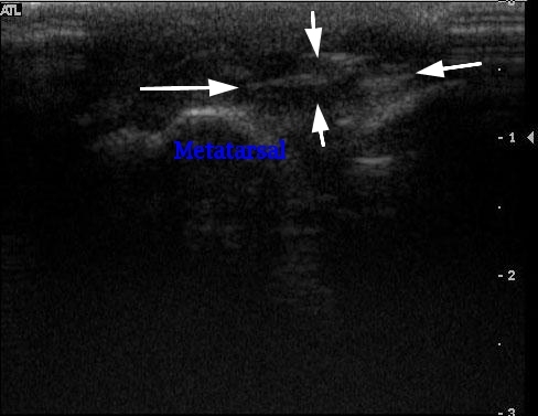
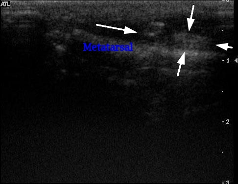
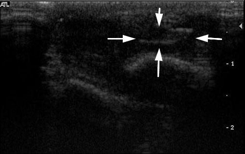
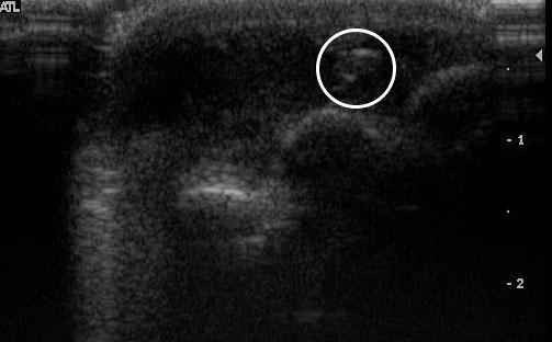
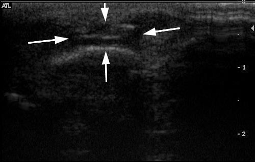
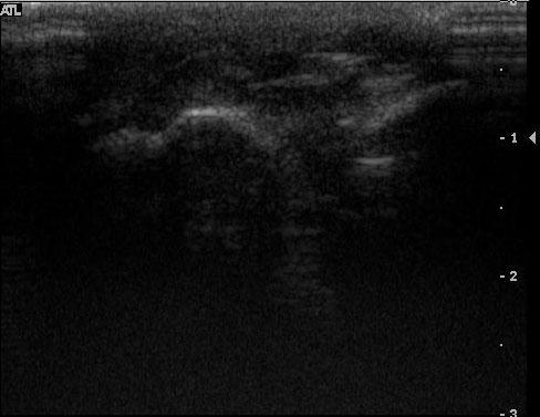
Today’s case comes straight from the ultrasound clinic. This is a 1 year old Pit Bull Terrier with a swelling over the 4th metatarsal bone of the left front limb for one month. Previous surgical exploration was unrewarding. Hint: click on the ‘Show annotated images’ link if you need help identifying the structures.

20090706010001_full_base 20090706010003_full_base 20090706010004_full_base 20090706010005_full_base 20090706010007_full_base

Show annotated images…

Annotated images

20090706010001_full_an 20090706010003_full_an 20090706010004_full_an 20090706010005_full_an 20090706010007_full_an

Show findings...

Findings

There is a similar appearing hyperechoic diamond shaped foreign body present between the fourth and fifth left metacarpal bones, oriented obliquely to the axis of the metacarpal bones. There is a large surrounding granuloma composed of hypoechoic tissue. There is no evidence of gas within the tissues.

Show diagnosis…

Diagnosis

Foreign body in the subcutaneous tissues of the left front foot, most likely a plant origin.

Show discussion…

Discussion

Plant foreign bodies can be seen as angular, hyperechoic structures within draining tracts or abscesses. In this case, there was no visible gas in a draining tract. The foreign body was clearly visible superficial to the 4th metatarsal bone.

Show additional image…

Additional image

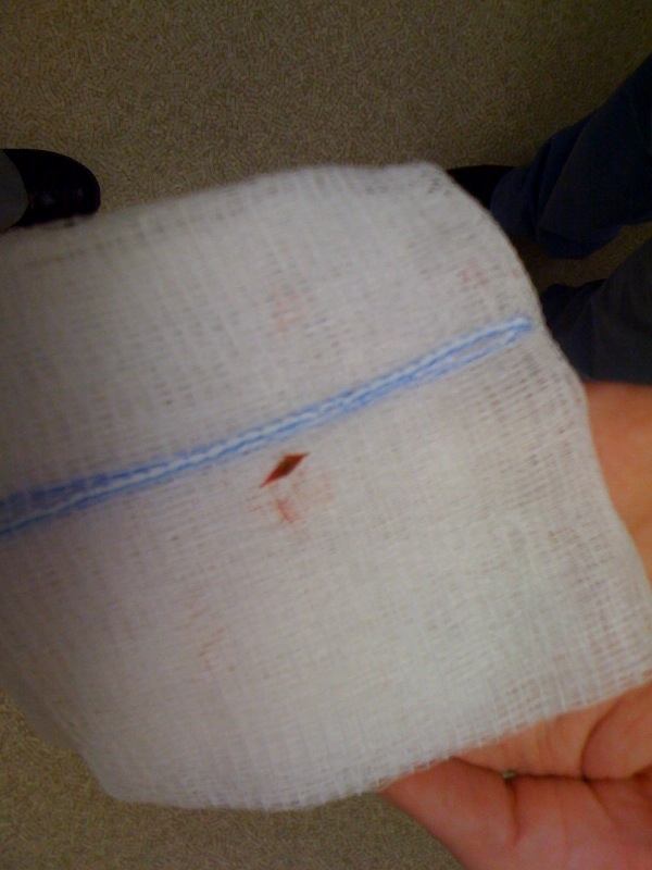
Here is the foxtail about 20 minutes after the ultrasound examination.

foxtail

foxtail
Image 1 of 1


Show references…

References

  1. Gnudi G, Volta A, Bonazzi M, et al. Ultrasonographic features of grass awn migration in the dog. Veterinary Radiology & Ultrasound 2005;46:423-426.
  2. Della Santa D, Rossi F, Carlucci F, et al. Ultrasound-guided retrieval of plant awns. Vet Radiol Ultrasound 2008;49:484-486.
  3. Armbrust LJ, Biller DS, Radlinsky MG, et al. Ultrasonographic diagnosis of foreign bodies associated with chronic draining tracts and abscesses in dogs. Veterinary Radiology & Ultrasound 2003;44:66-70.


Case originally posted on July 9, 2009
Filed Under: Canine, Case of the Day, Musculoskeletal, Ultrasound

  • Bloglovin
  • Facebook
  • RSS
  • Twitter
AtlasCover787x1024 amazon-availablenow

Veterinary Radiology News

Sign up for alerts about new cases and newsletters.

We will respect your privacy.

Archives

Recent Comments

Tags

abscess adenocarcinoma bronchiectasis carcinoma cardiomyopathy coccidioidomycosis discospondylitis elbow dysplasia eosinophilic bronchopneumopathy erosive polyarthritis feline infectious peritonitis FIP foreign body fracture gastric dilation gastric foreign body gastrointestinal GDV heart failure hemangiosarcoma hiatal hernia histiocytic sarcoma hypertrophic cardiomyopathy intestinal foreign body linear foreign body lymphoma megaesophagus osteochondrosis osteomyelitis osteosarcoma patent ductus arteriosus PDA pericardial effusion pneumonia pneumothorax polyarthritis PPDH pulmonary pulmonary abscess pulmonary adenocarcinoma pulmonary carcinoma sarcoma thymoma tracheal collapse vascular ring anomaly

Copyright © 2025 · Metro Pro Theme On Genesis Framework · WordPress · Log in