• Home
  • About
  • Articles
  • Books
  • Contact

Veterinary Radiology

Teaching and learning about veterinary diagnostic imaging.

  • Modality
    • Radiographs
    • Ultrasound
    • CT/MRI
  • Region
    • Thorax
    • Abdomen
    • Musculoskeletal
    • Neurologic
  • Species
    • Canine
    • Feline
    • Equine
    • Exotic

9 year old West Highland White Terrier

April 27, 2015 By Allison Zwingenberger

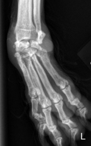
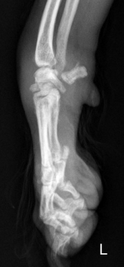
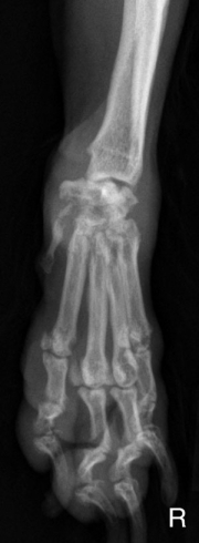
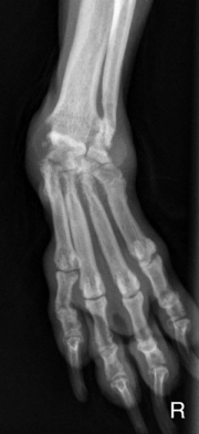
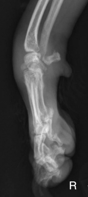
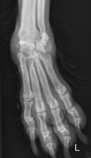
This is a great case; you don’t see one every day. It’s a 9 year old female neutered West Highland White Terrier with progressive reluctance to walk and abnormal gait. Her pelvic limbs are stiff and there is valgus of both carpi.

LF1
LF2
LF3
RF1
RF2
RF3

Show findings...

Findings

There is destruction of the subchondral bone of all proximal metacarpal bones, as well as the intermedioradial and second carpal bones. The third carpal bone is subluxated dorsally in both carpi, and the intermedioradial carpal bone is subluxated medially in the right carpus. There is soft tissue surrounding the carpus, and the carpal valgus is evident. The subluxation and angular deformity suggest instability of the joints. There are osteophytes on the doral aspect of both accessory carpal bones.

Show diagnosis…

Diagnosis

Erosive polyarthritis. The synovial fluid was purulent polyarthritis with 45-69% neutrophils and no organisms.

Show discussion…

Discussion

Erosive polyarthritis, or rheumatoid arthritis, causes inflammation of the synovium in an autoimmune pathway. The toxic byproducts of this inflammatory tissue attack the cartilage and subchondral bone in the joint. This excessive granulomatous tissue is called pannus and has the capacity to destroy articular cartilage, subchondral bone, and tendons of the affected joint. Proteases, enzymes, and other substances are produced by the invading tissue and are directly responsible for the destruction of the joint. In some cases, the pannus will grow beneath the articular cartilage, resulting in subchondral bone destruction. Articular cartilage loss begins at the joint margins. Radiolucent areas are frequently seen at the juxta-articular attachment of ligaments as well. As the condition progresses, loss of articular cartilage and subchondral bone continues, frequently resulting in a collapse or narrowing of the joint space.
Radiographic signs could include:

  • periarticular soft tissue swelling, multiple joints, bilaterally symmetrical (most commonly carpus and tarsus)
  • joint capsule effusion
  • joint space widening and later joint space narrowing
  • loss of subchondral and juxta-articular bone
  • subluxation, luxation, ankylosis of the affected joint
  • soft tissue atrophy, periarticular soft tissue mineralization
  • changes characteristic of degenerative joint disease

Show references…

References

  1. Carter SD, Barnes A, Gilmore WH. Canine rheumatoid arthritis and inflammatory cytokines. Veterinary Immunology and Immunopathology 1999;69:201-214.
  2. Rondeau MP, Walton RM, Bissett S, et al. Suppurative, nonseptic polyarthropathy in dogs. J Vet Intern Med 2005;19:654-662.
  3. Woodard JC, Riser WH, Bloomberg MS, et al. Erosive polyarthritis in two greyhounds. J Am Vet Med Assoc 1991;198:873-876.
  4. Pedersen NC, Castles JJ, Weisner K. Noninfectious canine arthritis: rheumatoid arthritis. J Am Vet Med Assoc 1976;169:295-303.


Case originally posted on May 11, 2007
Filed Under: Canine, Case of the Day, Musculoskeletal, Radiographs

  • Bloglovin
  • Facebook
  • RSS
  • Twitter
AtlasCover787x1024 amazon-availablenow

Veterinary Radiology News

Sign up for alerts about new cases and newsletters.

We will respect your privacy.

Archives

Recent Comments

Tags

abscess adenocarcinoma bronchiectasis carcinoma cardiomyopathy coccidioidomycosis discospondylitis elbow dysplasia eosinophilic bronchopneumopathy erosive polyarthritis feline infectious peritonitis FIP foreign body fracture gastric dilation gastric foreign body gastrointestinal GDV heart failure hemangiosarcoma hiatal hernia histiocytic sarcoma hypertrophic cardiomyopathy intestinal foreign body linear foreign body lymphoma megaesophagus osteochondrosis osteomyelitis osteosarcoma patent ductus arteriosus PDA pericardial effusion pneumonia pneumothorax polyarthritis PPDH pulmonary pulmonary abscess pulmonary adenocarcinoma pulmonary carcinoma sarcoma thymoma tracheal collapse vascular ring anomaly

Copyright © 2025 · Metro Pro Theme On Genesis Framework · WordPress · Log in