• Home
  • About
  • Articles
  • Books
  • Contact

Veterinary Radiology

Teaching and learning about veterinary diagnostic imaging.

  • Modality
    • Radiographs
    • Ultrasound
    • CT/MRI
  • Region
    • Thorax
    • Abdomen
    • Musculoskeletal
    • Neurologic
  • Species
    • Canine
    • Feline
    • Equine
    • Exotic

6 year old German Shepherd

March 2, 2015 By Allison Zwingenberger

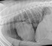
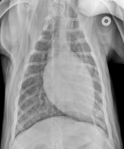
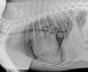
Today’s case is a 6 year old male neutered German Shepherd with anorexia. He was diagnosed with renal failure at the emergency veterinary clinic and referred for diagnostics and treatment. Post your interpretations in the comments!

L LAT Thorax
R LAT Thorax
DV Thorax

Show findings...

Findings

There is diffuse unstructured interstitial pattern in the lung parenchyma, most apparent in the caudodorsal region. The cardiovascular structures appear normal. There is also an intravenous catheter visible in the caudal vena cava.

Show differential diagnosis…

Differential Diagnosis

  • Pneumonitis (metabolic due to uremia)
  • Non-cardiogenic pulmonary edema
  • Hemorrhage
  • ARDS/SRRS

Show diagnosis…

Diagnosis

  • Uremic pneumonitis secondary to renal failure.

Case originally posted on May 22, 2009
Filed Under: Canine, Case of the Day, Radiographs, Thorax

Comments

  1. vet81 says

    May 22, 2009 at 1:18 pm

    There is a diffuse peribronchial and interstitial lung pattern in the middle and caudal lung lobes on both sides. The is a small amount of fluid in the pleural cavity. The heart silhouette is without normal limits. Lung vessels seem to be widened. It could be a pneumonia due to chemical intoxication.

  2. Vet76 says

    June 4, 2009 at 9:24 pm

    The answers are already there but I tried to interpret the images before I read your comments. So here it is. I thought that the predominant pattern was interstitial although has also a alveolar pattern. Definitively the caudo dorsal lung lobes were more affected. Given the history of acute renal failure the first thought that came to my mind was fluid overload vs PLN with secondary leakage of fluids. Pneumonitis was not in my differentials (so good thing I read your interpretation). In retrospective (after I read your interpretation). Pulmonary vessels appeared full but not severely enlarged( potentially ruling out fluid overload) and Leakage usually occurs in pleural space or peritoneal space (which do not appear to be present in significant amounts). I have two questions do you have a recent article that I can read about pneumonitis specifically uremic pneumonitis, and also is there also information about the technique for vena cava catheterization ( does it have an proper name ?). Thank you again

    • Allison Zwingenberger says

      June 5, 2009 at 1:56 pm

      Those are good observations. Textbooks might be the most helpful source of information on pneumonitis, such as the BSAVA manual of thoracic imaging listed under the resources section.

  • Bloglovin
  • Facebook
  • RSS
  • Twitter
AtlasCover787x1024 amazon-availablenow

Veterinary Radiology News

Sign up for alerts about new cases and newsletters.

We will respect your privacy.

Archives

Recent Comments

Tags

abscess adenocarcinoma bronchiectasis carcinoma cardiomyopathy coccidioidomycosis discospondylitis elbow dysplasia eosinophilic bronchopneumopathy erosive polyarthritis feline infectious peritonitis FIP foreign body fracture gastric dilation gastric foreign body gastrointestinal GDV heart failure hemangiosarcoma hiatal hernia histiocytic sarcoma hypertrophic cardiomyopathy intestinal foreign body linear foreign body lymphoma megaesophagus osteochondrosis osteomyelitis osteosarcoma patent ductus arteriosus PDA pericardial effusion pneumonia pneumothorax polyarthritis PPDH pulmonary pulmonary abscess pulmonary adenocarcinoma pulmonary carcinoma sarcoma thymoma tracheal collapse vascular ring anomaly

Copyright © 2025 · Metro Pro Theme On Genesis Framework · WordPress · Log in