Today’s case is a 4-year-old female neutered English Bull Dog with history of multiple cutaneous masses. Presents non-ambulatory with severe neck pain. Bloodwork shows hypercalcemia (14.5 mg/dL), leukocytosis (31,000/uL), and mild regenerative anemia (PCV 35%, reticulocytes 276,000/uL). Do you have any additional input or questions about the images? Post your comments here!
Findings
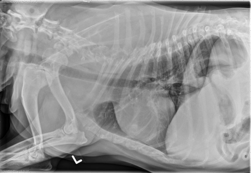
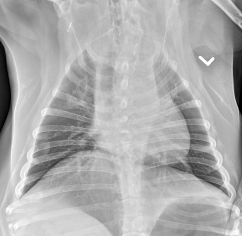
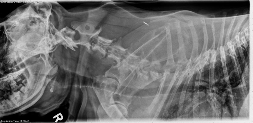
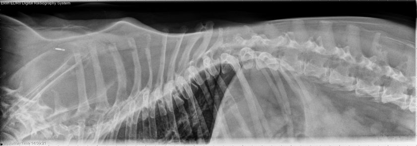
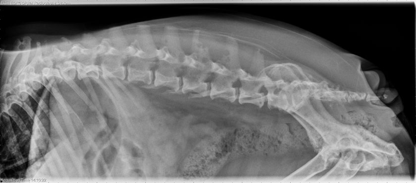
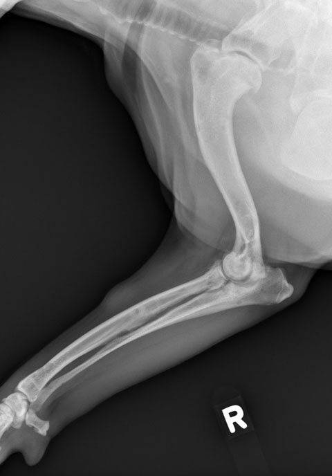
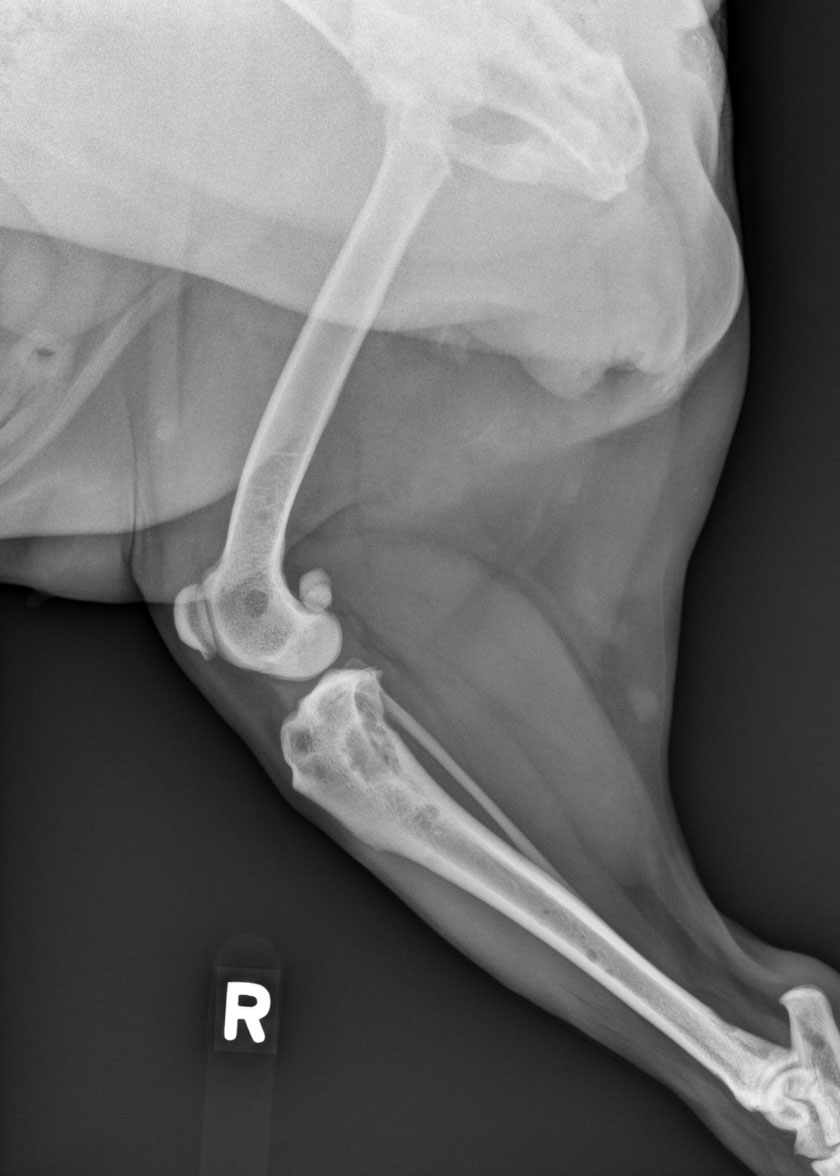
The structures of the thorax appear normal for the age and breed. However, multiple lytic areas are visible in the dorsal spinous processes on the lateral radiographs. These were confirmed on spine technique radiographs, where they are also seen to involve the vertebral bodies, scapula, pelvis and skull. The survey was expanded to include the forelimbs and hindlimbs. The long bones also have areas of lysis with a short transition zone. Several lesions are surrounded by rims of sclerotic bone.
Differential Diagnosis
- Metastatic neoplasia – hemangiosarcoma, osteosarcoma
- Multiple myeloma/plasma cell tumor
- Lymphosarcoma
Diagnosis
Cutaneous hemangiosarcoma with diffuse metastatic disease and multiple pathologic fractures of cervical vertebrae.
Discussion
This multifocal, aggressive pattern of lysis can be due to primary (round cell) or metastatic neoplasia as listed under differential diagnoses. A multifocal inflammatory lesion such as fungal osteomyelitis usually has more production associated with the lesions.