Happy New Year to all my VR readers! I hope you are feeling rested and recharged for the year ahead. On that note, enjoy this case! It is a 4-year-old Brittany Spaniel with one episode of vomiting, several incidences of diarrhea, anorexia, and lethargy for the last two days.
Findings
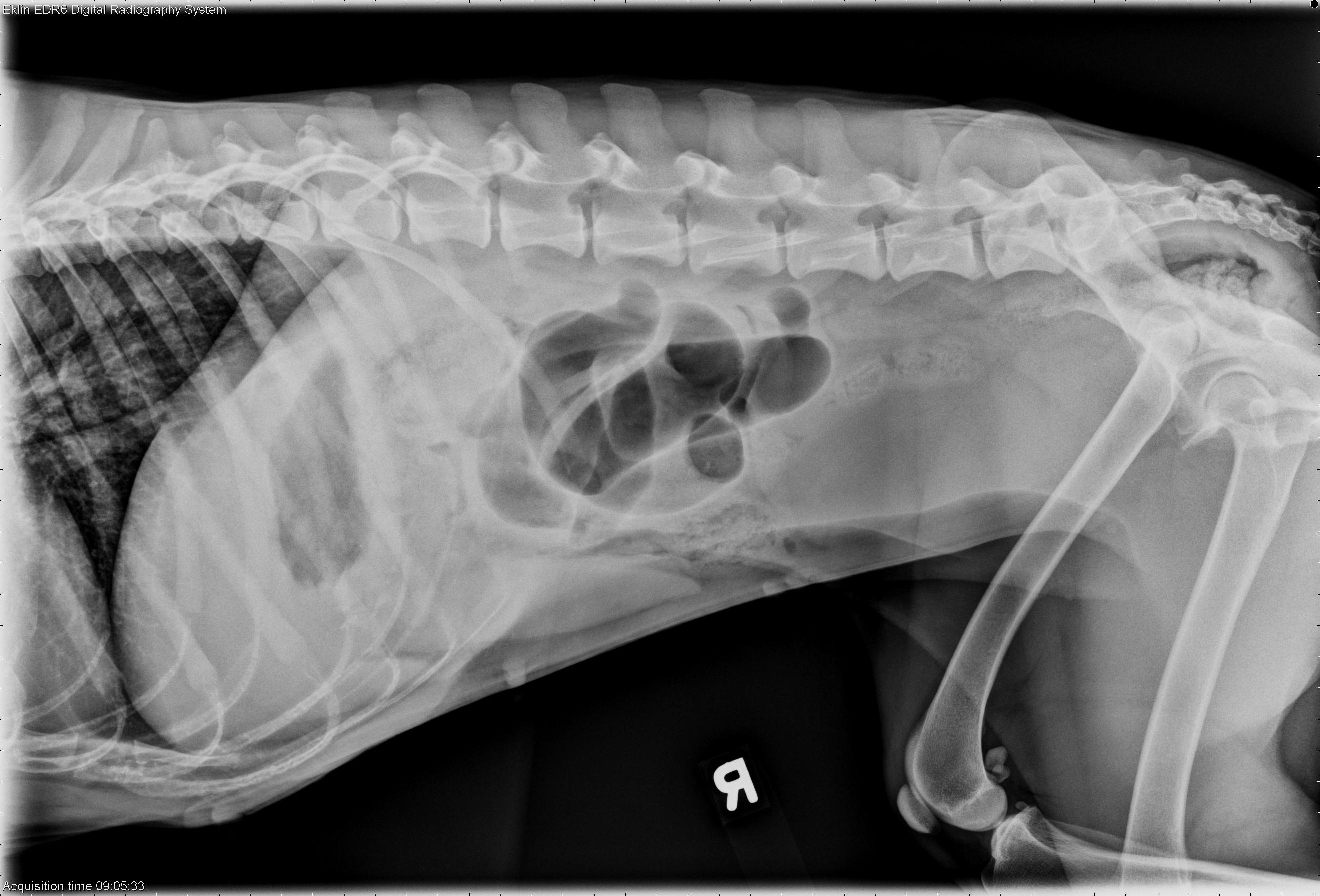
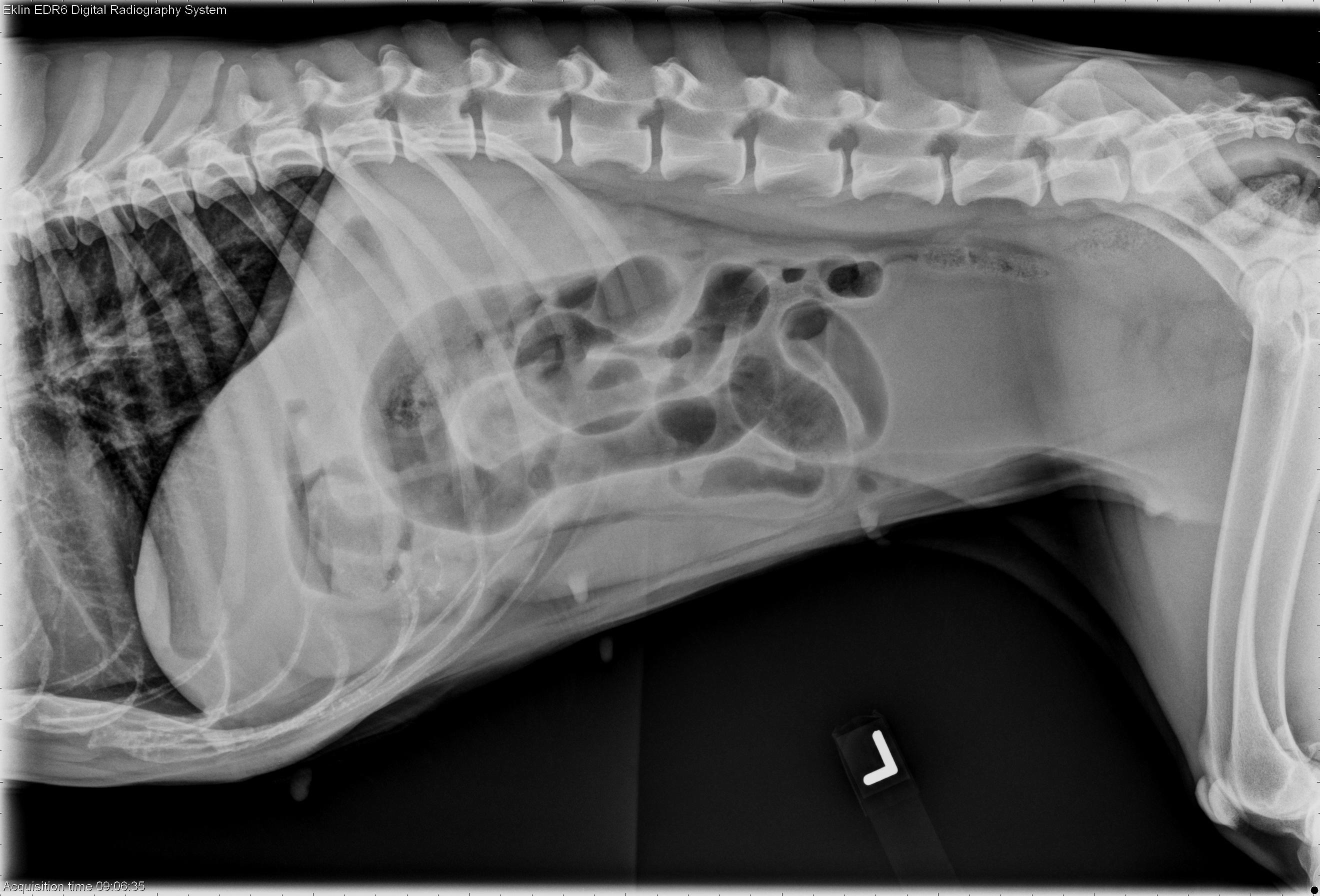
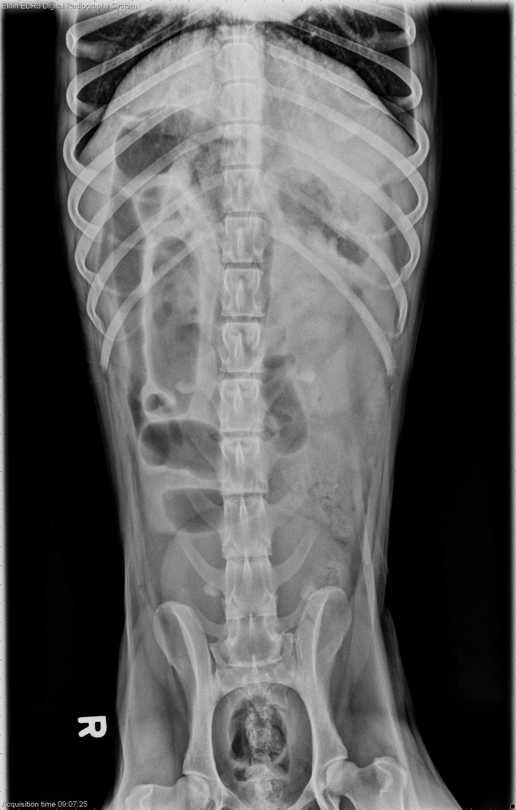
The duodenum plus additional small intestine are dilated with gas. On the right lateral projection, there are normally sized small intestinal loops visible.Additionally, there is soft tissue opacity foreign material in the pylorus on the left lateral projection. It is suspended in the pylorus and surrounded by air.
Differential Diagnosis
Small intestinal obstruction, most likely due to foreign bodyDiagnosis
Foreign body in stomach and jejunum – corn cobDiscussion
The foreign material in the stomach was indicative of a foreign body obstruction. The dilated small intestine was due to the more distal jejunal piece of corn cob. Pyloric obstruction often results in a more dilated and fluid filled stomach, except if the animal has been vomiting, or there is a partial obstruction.The key to diagnosing this case is in observing the two distinct populations of small intestine; enlarged greater than 1.5x the height of a vertebral body, and more normal appearing intestine.
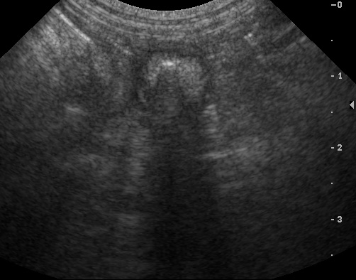
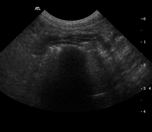
Ultrasound images confirm the presence of “hard shadowing”, or attenuating, foreign material in the small intestine. Gas generally produces “dirty” shadowing, or multiple reverberation artifacts.
References
- Tidwell AS, Penninck DG. Ultrasonography of gastrointestinal foreign bodies. Veterinary Radiology 1992;33:160-169.
- Boysen SR, Tidwell AS, Penninck DG.