• Home
  • About
  • Articles
  • Books
  • Contact

Veterinary Radiology

Teaching and learning about veterinary diagnostic imaging.

  • Modality
    • Radiographs
    • Ultrasound
    • CT/MRI
  • Region
    • Thorax
    • Abdomen
    • Musculoskeletal
    • Neurologic
  • Species
    • Canine
    • Feline
    • Equine
    • Exotic

4 year old Thoroughbred

August 17, 2015 By Allison Zwingenberger

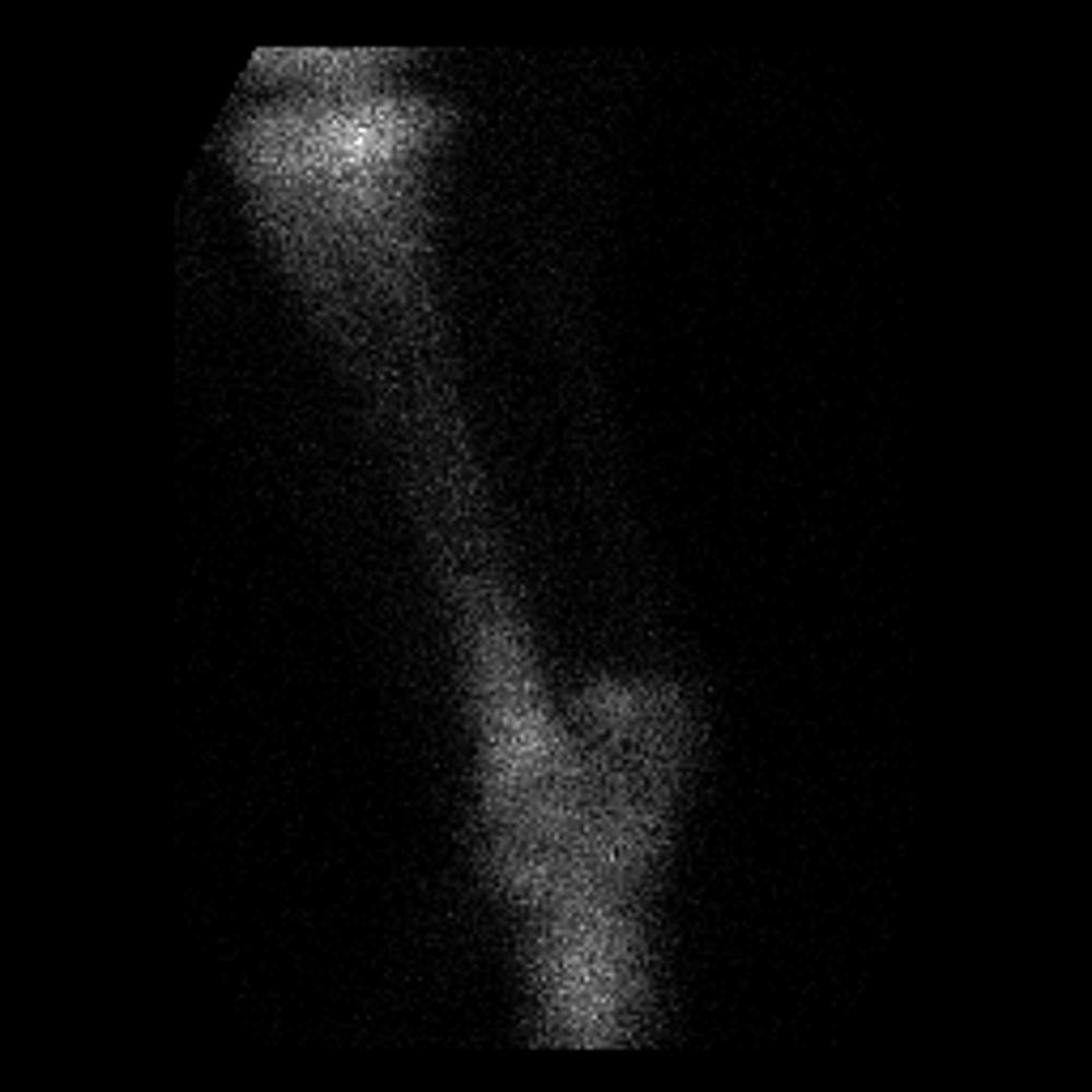
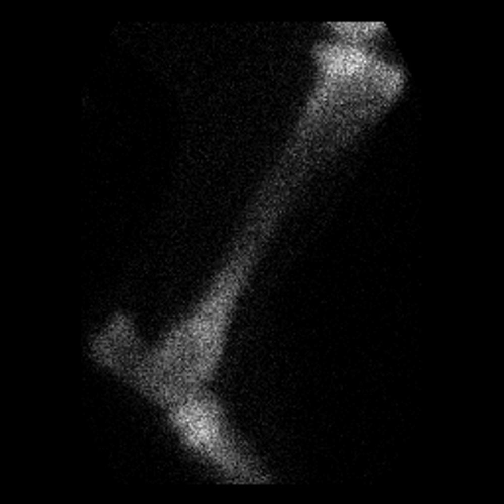
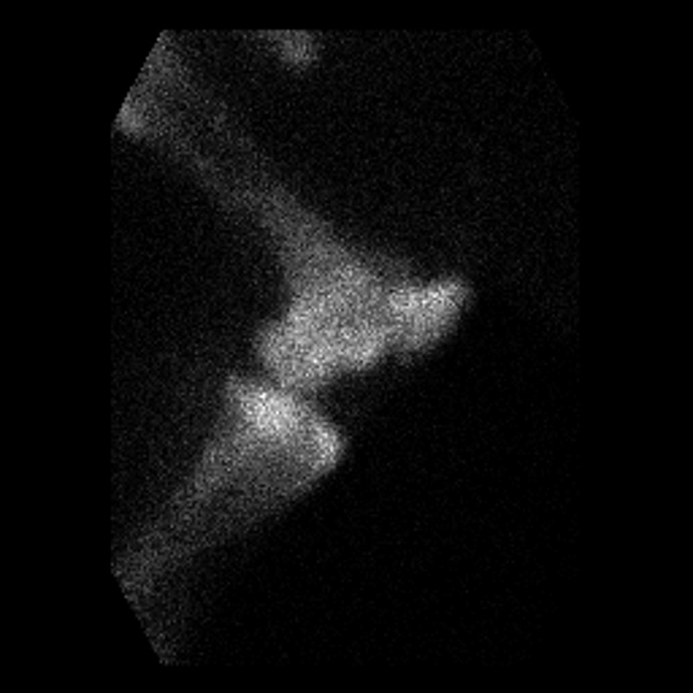
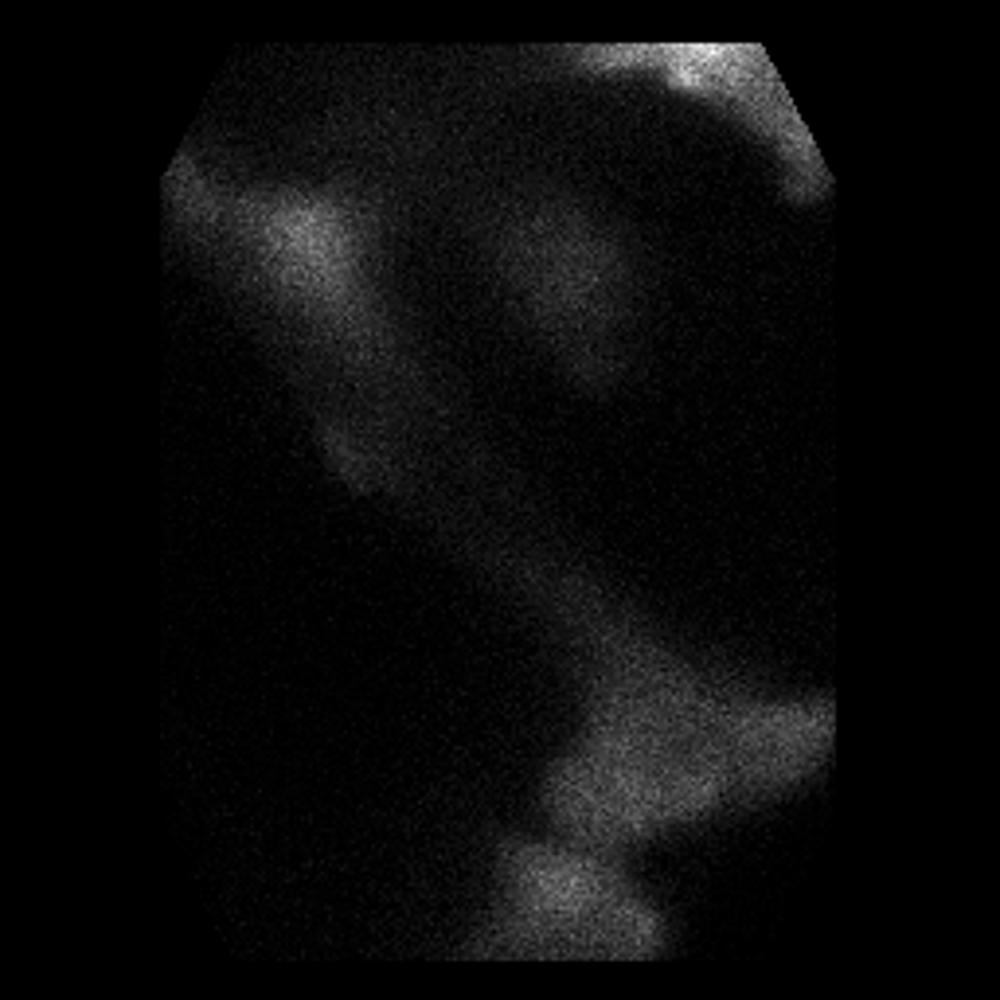
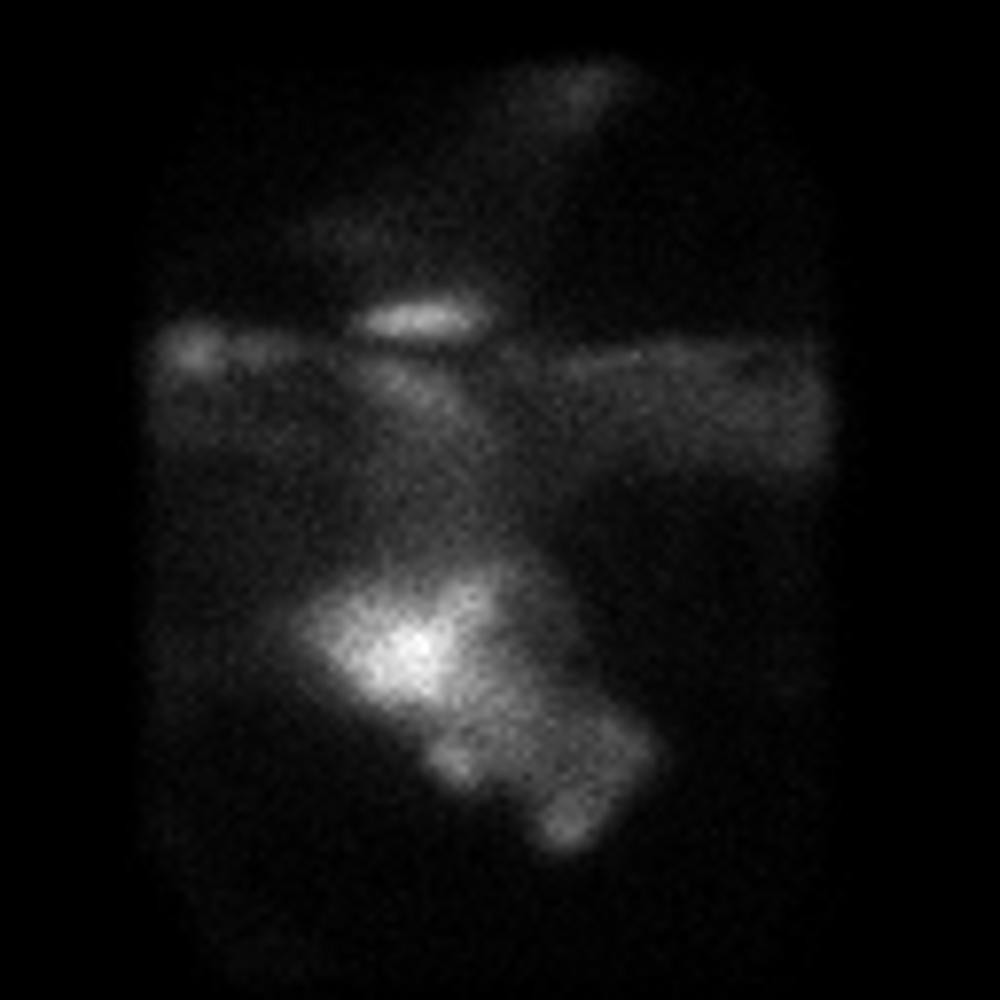
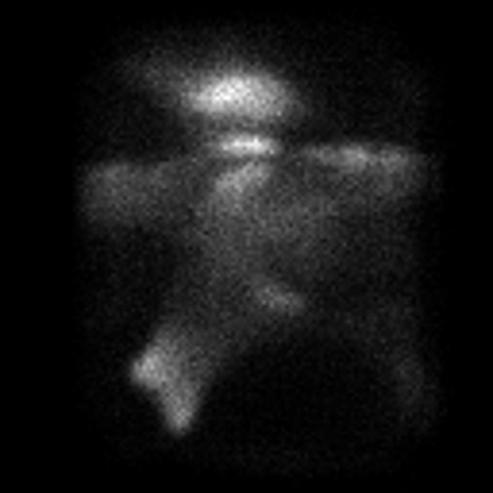
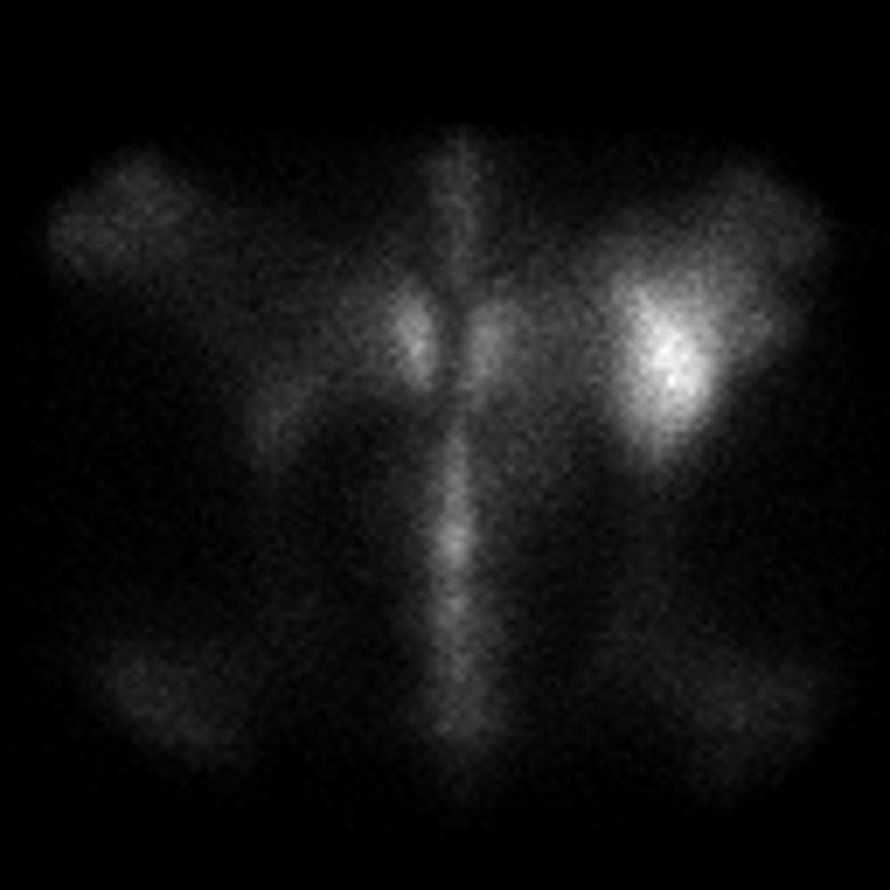
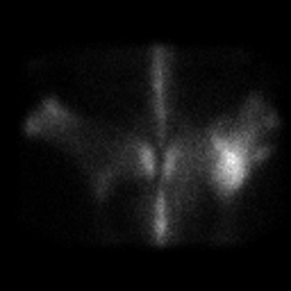
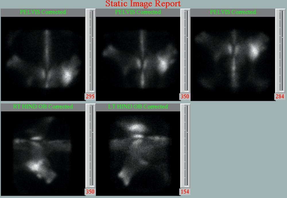
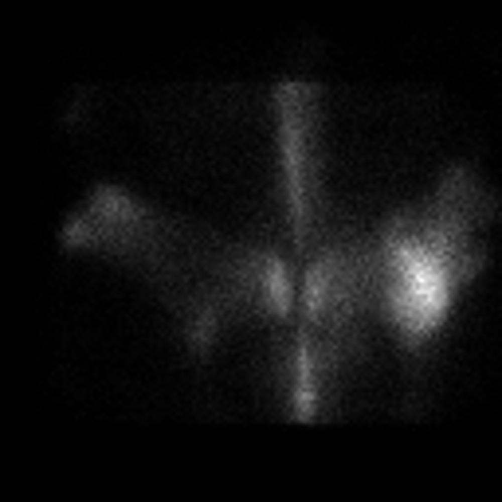
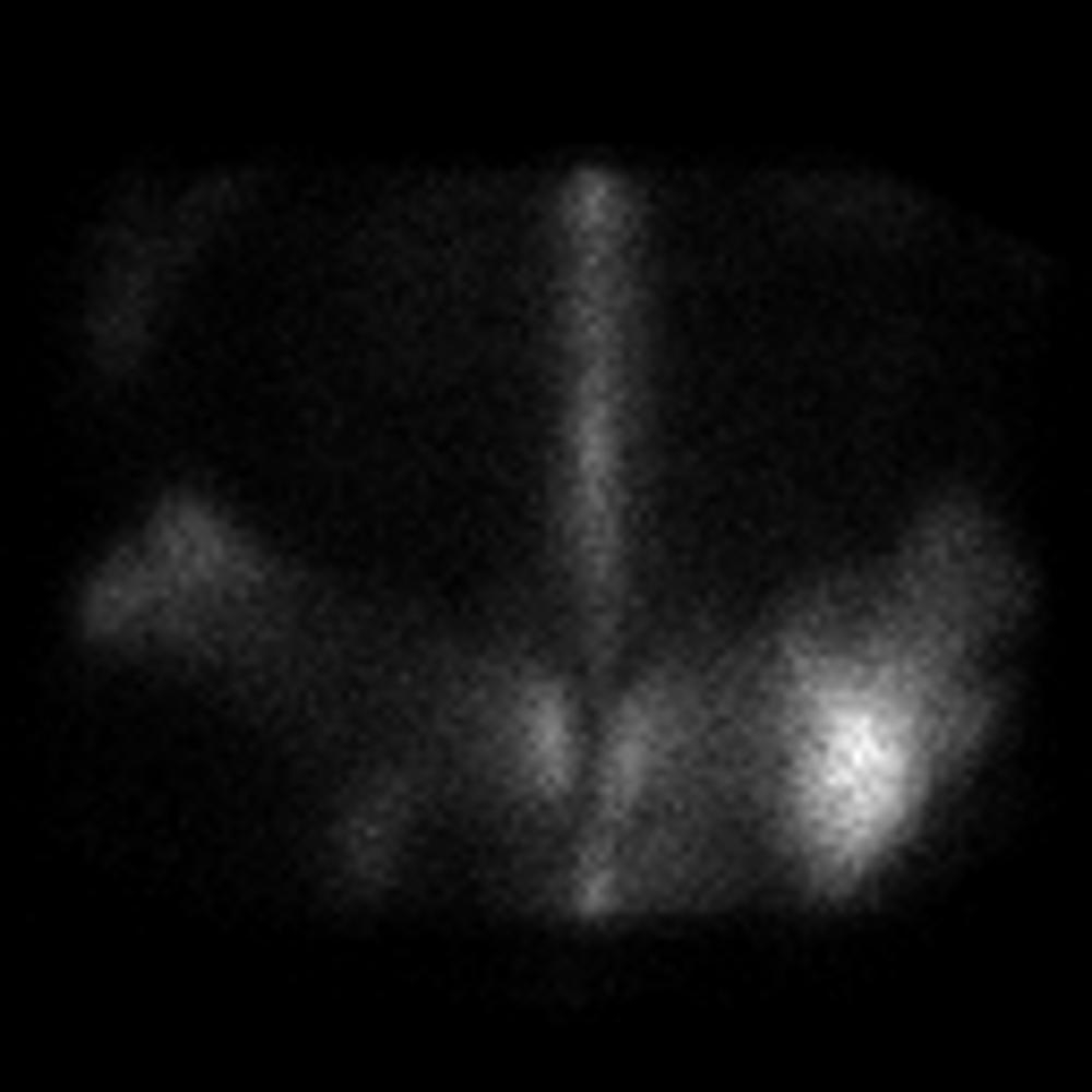
Today’s case takes you into the world of equine nuclear scintigraphy! For those of you that haven’t seen one before, we inject Technetium-99m-MDP, which is a radioisotope that localizes to active bone. After a few hours, we can take the images and find areas of increased uptake that can indicate inflammation, fracture, or neoplasia. The images will look grainy because they have low spatial resolution, but they are very sensitive to small changes in activity. This type of functional imaging can give us a lot of information about biological processes. Take a look!

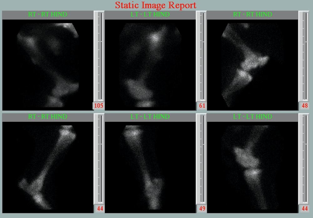
The case is a 4 year old Thoroughbred filly in race training with intermittent right hind limb lameness for the last few months.
Radiographs of the tarsus and tibia are normal.
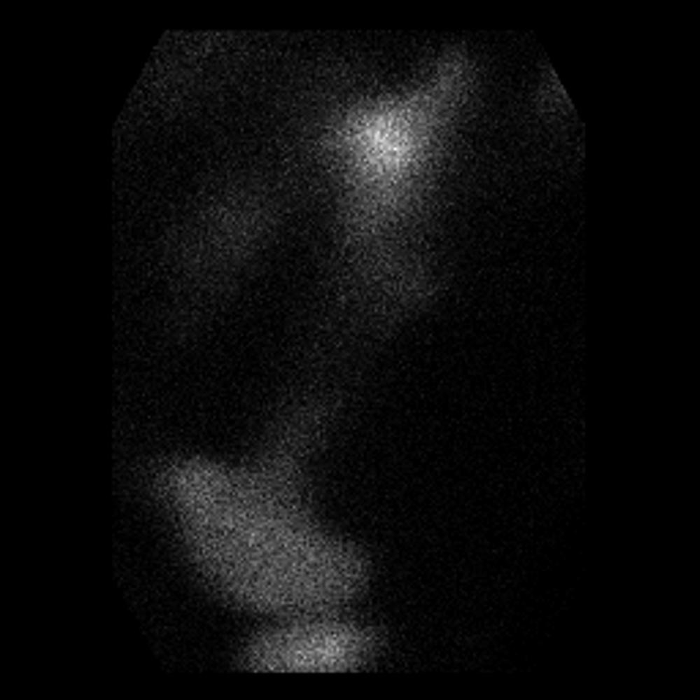
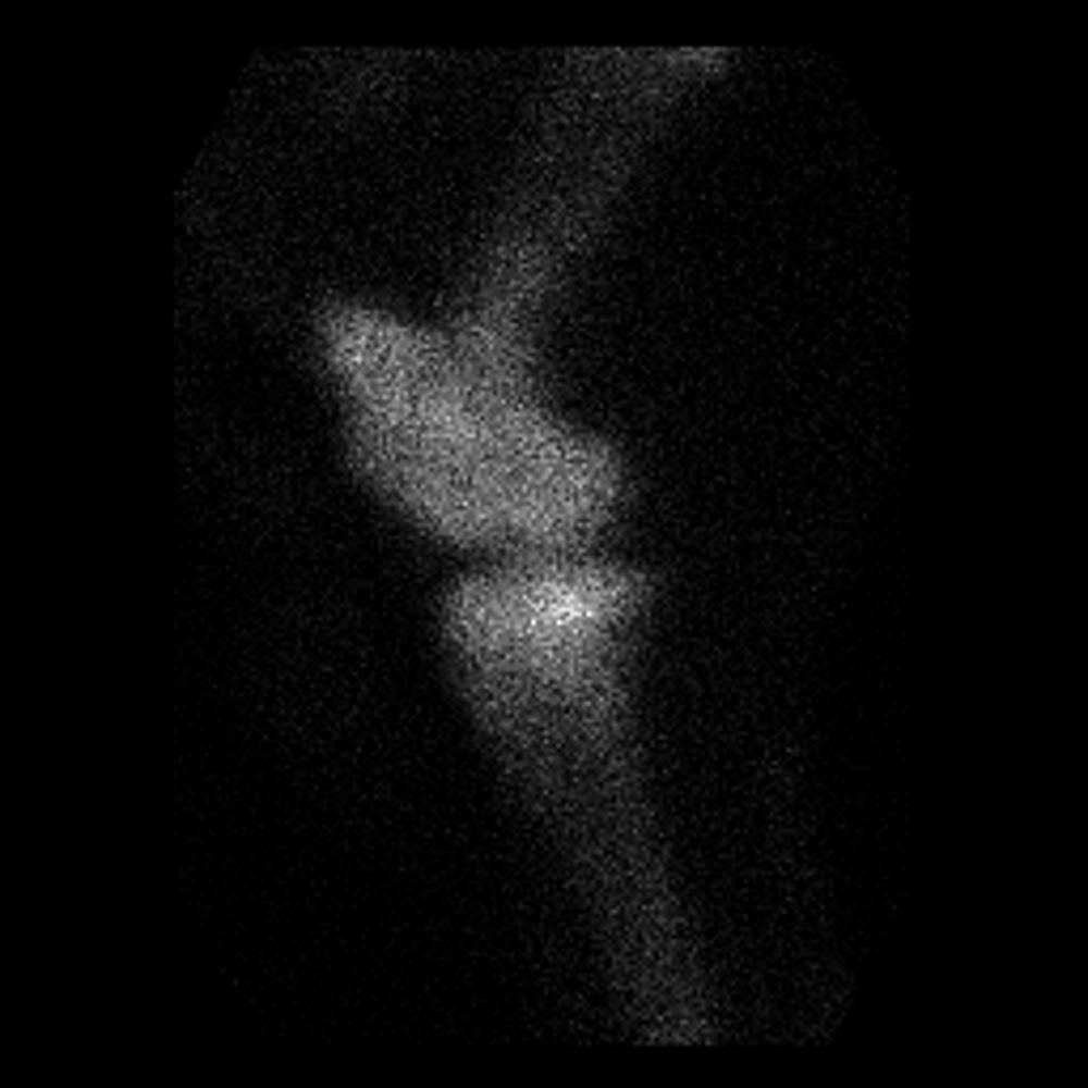
LT HIND LAT LT HIND LAT LT HIND LAT RT HIND LAT RT HIND RT RT HIND RT RT HIND OB Corrected LT HIND OB Corrected PELVIS Corrected PELVIS Corrected Static Image Report PELVIS Corrected PELVIS Corrected Static Image Report

Show findings...

Findings

There is increased uptake of radioisotope on the right ilium, which indicates focal bone remodeling. The hind limbs are normal.

Show diagnosis…

Diagnosis

Stress fracture of the right ilium.

Show discussion…

Discussion

Common areas for stress fractures in racehorses include the tibia, humerus, scapula, metacarpus, and ilium. Oblique views of the pelvis, in addition to dorsal views, can help to isolate the lesion.
Hornof, W. J., S. M. Stover, P. D. Koblik and R. M. Arthur (1996). “Oblique views of the ilium and the scintigraphic appearance of stress fractures of the ilium.” Equine Veterinary Journal 28(5): 355-358.
Mackey, V. S., D. R. Trout, D. M. Meagher and W. J. Hornof (1987). “Stress fractures of the humerus, radius, and tibia in horses. Clinical features and radiographic and/or scintigraphic appearance.” Veterinary Radiology 28(1): 26-31.

Case originally posted on January 29, 2009

Filed Under: Case of the Day, Equine, Musculoskeletal

  • Bloglovin
  • Facebook
  • RSS
  • Twitter
AtlasCover787x1024 amazon-availablenow

Veterinary Radiology News

Sign up for alerts about new cases and newsletters.

We will respect your privacy.

Archives

Recent Comments

Tags

abscess adenocarcinoma bronchiectasis carcinoma cardiomyopathy coccidioidomycosis discospondylitis elbow dysplasia eosinophilic bronchopneumopathy erosive polyarthritis feline infectious peritonitis FIP foreign body fracture gastric dilation gastric foreign body gastrointestinal GDV heart failure hemangiosarcoma hiatal hernia histiocytic sarcoma hypertrophic cardiomyopathy intestinal foreign body linear foreign body lymphoma megaesophagus osteochondrosis osteomyelitis osteosarcoma patent ductus arteriosus PDA pericardial effusion pneumonia pneumothorax polyarthritis PPDH pulmonary pulmonary abscess pulmonary adenocarcinoma pulmonary carcinoma sarcoma thymoma tracheal collapse vascular ring anomaly

Copyright © 2023 · Metro Pro Theme On Genesis Framework · WordPress · Log in