One evening a week, our group gets together to read known cases. That is, they are known to the faculty member who brought them, but unknown to everyone else. It’s a forum for the radiology residents to practice oral case reading style, and to see lots of cool images. We get a great turnout, from all the residents, any visitors that happen to be here, private practice radiologists and private practitioners, and even the emeriti faculty. It’s an awesome font of veterinary radiological knowledge in one room.
Usually one resident has one case to read, then the other residents add any other findings, and the faculty give their 2 cents (more like a dollar). In the discussion about the case, before and after the diagnosis is revealed, there are often pearls of knowledge that come to the surface, and everyone learns something.
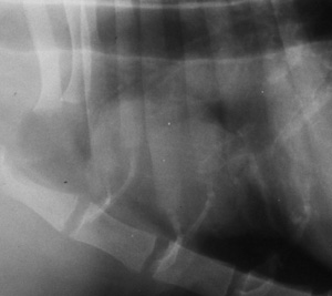
This week we looked at equine radiographs of the skull, thorax, and limbs. There was one case that had a large soft tissue mass in the thorax, that was perfectly clearly circumscribed by air, with no surrounding lung pathology. Differentials that came out were pulmonary mass, mediastinal mass and pleural mass with pneumothorax. It turned out to be a lymphoma originating from the heart base; not a common equine condition. But one comment that stuck with me was that a pulmonary mass of inflammatory origin should have some sort of pulmonary reaction surrounding it, like an interstitial pattern. Masses that look perfectly smooth and well-defined tend to be neoplastic, or benign such as a cyst, and don’t cause local lung inflammation. A second comment was that it didn’t contain any air, and that masses of pulmonary origin usually do. Both of these opinions held up in this case, supporting the non-pulmonary and non-inflammatory nature of the mass.