• Home
  • About
  • Articles
  • Books
  • Contact

Veterinary Radiology

Teaching and learning about veterinary diagnostic imaging.

  • Modality
    • Radiographs
    • Ultrasound
    • CT/MRI
  • Region
    • Thorax
    • Abdomen
    • Musculoskeletal
    • Neurologic
  • Species
    • Canine
    • Feline
    • Equine
    • Exotic

21 year old Scarlet Macaw

December 21, 2015 By Allison Zwingenberger

This week we have an avian case contributed by Dr. Leila Marcucci of the Bay Area Bird Hospital in San Francisco. It’s a 21-year-old female Scarlet Macaw who presented for increased water intake, watery droppings, and decreased appetite. Have a look and post your comments below.

R LAT Abdomen
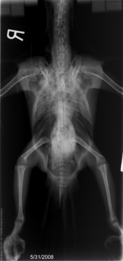
VD Abdomen

Show findings...

Findings

5/31/08 – The lateral radiograph demonstrated a 4.5 cm diameter, well-circumscribed, soft tissue opacity in the central abdominal area. It extended from the caudal hepatic border to the caudal pelvic region. Its dorsal margin was relatively distinct and did not silhouette with the kidneys. Evidence of calcification was not identified. Increased endosteal opacity was present in the pelvic extremities. The remainder of the study was unremarkable except for mild decreased liver size.

Show differential diagnosis…

Differential Diagnosis

  • Soft tissue opacity mass – retained egg (within the reproductive tract vs. free in the coelom), less likely splenic mass.
  • Endosteal opacity – indicates reproductive activity.

Show followup…

Followup

5/31/08 – Conservative medical management was selected for the retained egg. Treatment consisted of calcium lactate/clacium glycerpohospate (Calphosan, l5 mg/ml, 1 cc IM) IM Injection and a gavage of a commercial avian diet. The bird was discharged for home care.
6/2/08 – Represented the following Monday (June 2). In an attempt to medically address the retained egg, the bird was hopsitalized and given fluids (LRS-W 35 cc SQ), IM calcium (Calphosan, l5 mg/ml, 1 cc IM) and butorphanol (10 mg/ml 0.32 cc IM). Bloodwork taken on 5/31 showed elevations in GGT (9 U/L), amylase (1024 U/L), glucose (379 mg/dl), uric acid (15.3 g/dl) and WBC (43,000). WBC morphology was within normal limits although there was an increased percentage of heterophils and a decreased percentage of lympocytes. A salpingitis or coelomitis was suspected and the bird was started on injectable antibiotics (Cefotaxime 200 mg/ml 0.5 cc IM). While hospitalized, the bird’s appetite was decreased, so she was gavaged twice daily.


R LAT Abdomen-Followup

6/3/08 – A recheck lateral radiographic study made on 6/3/08 and demonstrated persistence of the mass effect. Its margination was somewhat less distinct than on the previous studies, but individual intestinal segments could be identified indicating a large amount of free coelomic fluid was not present. No evidence of pulmonary consolidative disease or cardiac enlargement was identified.

ultrasound.0001
ultrasound.0002

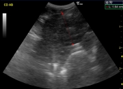
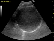
Coelomic ultrasound was performed. A 3 cm rounded hypoechoic homogeneous mass effect occupied the caudal coelom.It had a thin uniform wall. There was no evidence of free coelomic fluid. It was concluded that the the rounded mass was a non viable egg, free within the coelomic cavity.

Show diagnosis…

Diagnosis

The following day (June 4) the bird was taken to surgery for egg removal. Coelomic exploration revealed extremely thickened peritoneal membranes and a soft-shelled egg free in the coelom. The egg was easily removed and a salpingohysterectomy performed. The bird awoke from the surgery without incident. The following day she was vocal, active and eating well on her own.
The soft-shelled egg was filled with a foul smelling liquid. Histopathology revealed inflammatory cells present on the surface including multinucleate giant cells, lymphocytes and plasma cells. Although the oviduct appeared to have a stricture in the submucosa on gross inspection, no lesions were noted on histopathology.

R LAT Abdomen-Retained Egg

Show discussion…

Discussion

The initial radiographs were unusual because of the distinct margination of the egg in the coelom. Reproductive masses and free coelomic eggs are often poorly marginated, either by being surrounded by reproductive tissues or fluid from yolk coelomitis.
One week post surgery the white cell count was back in the normal range (12,000). By July 2008, the bird appeared normal, the incision was fully healed and her weight had returned to a more normal range (1115 gm)

Case originally posted on November 27, 2008

Filed Under: Abdomen, Case of the Day, Exotic, Radiographs, Ultrasound

Comments

  1. vet74 says

    November 27, 2008 at 7:18 am

    The vertebras and the areas of the femoral head and humeral head seem a more radiolucent than what I would expect, but on the other hand I haven’t seen many rads of Macaws…

  2. giglior says

    November 27, 2008 at 5:48 pm

    Hi there!

    There is a rounded, well defined, soft tissue opaque mass in the caudal coelom, superimposed to the pelvic limbs. I think the hepatic silhouette is decreased in size. There a heterogeneous increased opacity within the medulary cavities of the femur and tibiotarsus, bilaterally. My first differential for that is polyostotic hyperostosis with a non-calcified egg. The possibility of a coelomic neoplasia such as oviductal tumor also needs to be considered.
    Have an excellent Thanksgiving!!

  3. Sam Verstraete says

    December 8, 2008 at 7:29 am

    Hi there,

    as a therapy they performed a salpingohysterectomy. Why didn’t they removed the ovarium. What happens with the next ege. It will fall in the abdomen cavity and caused an ege-peritonitis?

  4. Allison Zwingenberger says

    December 9, 2008 at 3:13 pm

    Sam, here’s the response from Dr. Marucci:

    A salpyngohysterectomy does not include removal of the ovaries – they are intimately associated with the aorta and attempted surgical removal has a high risk of severe hemorrhage.

    The concern is that follicles will mature and drop into the coelomic cavity, possibly causing an egg yolk coelomitis. This is a valid concern and is a side effect we often see. It is possible for birds to drop an active follicle and not generate an infection or only develop a mild self-limiting infection. In this bird’s case, she has a history of only 1 egg being produced previously. She has had a single incident of egg yolk coelomitis in the past (with her reproductive tract intact!). Therefore, she has a much lower risk of egg yolk coelomitis than some birds.

  • Bloglovin
  • Facebook
  • RSS
  • Twitter
AtlasCover787x1024 amazon-availablenow

Veterinary Radiology News

Sign up for alerts about new cases and newsletters.

We will respect your privacy.

Archives

Recent Comments

Tags

abscess adenocarcinoma bronchiectasis carcinoma cardiomyopathy coccidioidomycosis discospondylitis elbow dysplasia eosinophilic bronchopneumopathy erosive polyarthritis feline infectious peritonitis FIP foreign body fracture gastric dilation gastric foreign body gastrointestinal GDV heart failure hemangiosarcoma hiatal hernia histiocytic sarcoma hypertrophic cardiomyopathy intestinal foreign body linear foreign body lymphoma megaesophagus osteochondrosis osteomyelitis osteosarcoma patent ductus arteriosus PDA pericardial effusion pneumonia pneumothorax polyarthritis PPDH pulmonary pulmonary abscess pulmonary adenocarcinoma pulmonary carcinoma sarcoma thymoma tracheal collapse vascular ring anomaly

Copyright © 2025 · Metro Pro Theme On Genesis Framework · WordPress · Log in