I hope everyone had a wonderful holiday season. We are planning an exciting facelift for VR to make the site more user friendly and accessible to mobile devices. The site will be in maintenance mode for the next few days as we make the upgrades, so check back later this week to see how it looks! … [Read more...]
CT/MRI Society Case of the Month
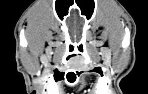
This month we are featuring a CT case authored by Elizabeth Ballegeer of Michigan State University. Enjoy the case! Access all case images … [Read more...]
How can Veterinary Radiology help you?
I love getting questions from VR readers because it helps me to find out more about what you'd like to learn about interpreting diagnostic images. To find out more, I've posted a survey where you can tell me exactly what you have problems with and what you'd like to accomplish. I'm looking forward … [Read more...]
Known Case Conference
11 year old male neutered German Shepherd with 5 days of lethargy and 1 day of vomiting. On abdominal radiographs, there is a large mid abdominal mass which is displacing small intestine to the periphery. The pylorus of the stomach is displaced slightly dorsally, and the fundus is caudally … [Read more...]
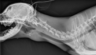
Finding The Origin Of A Thoracic Mass
This week at known case conference we had a collection of interesting thoracic cases. When evaluating masses, try to determine where they are originating and what surrounding structures they affect. There are some clues below that help with localization in each case. 4 year old German Shepherd with … [Read more...]
- 1
- 2
- 3
- …
- 8
- Next Page »
Recent Comments