I’ve been experimenting with upgrading my hardware and software ever since we got a 16 slice CT machine. Before that, whatever software I was using (eFilm, Osirix) seemed to do ok with the images. But the advent of the CT study with 1000, 2000, or 3000 images really challenged the average mac or … [Read more...]
CT/MRI Society Case of the Month – January 2013
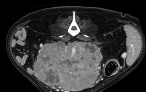
This month's case from the CT/MRI Society of the ACVR is a CT scan provided by Dr. Rob McLear. It has a holiday theme... … [Read more...]
CT/MRI Society Case of the Month – October 2012
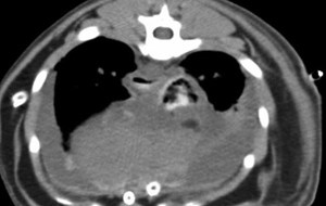
This month we have a case from Dr. Alex zur Linden of the University of Guelph. These cases are sponsored by the CT/MRI Society of the ACVR and are available to view for 1 month. For full access to the case archive, join the CT/MRI society at the ACVR website. Access all images and … [Read more...]
CT/MRI Society Case of the Month
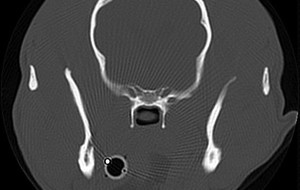
This month's CT/MRI Society case is presented by Jennifer Reetz, from the University of Pennsylvania. If you'd like unlimited access to all CT/MRI society cases, please join the society at the ACVR website. Cases provided here are available to the public for viewing for a period of one … [Read more...]
CT/MRI Society Case of the Month
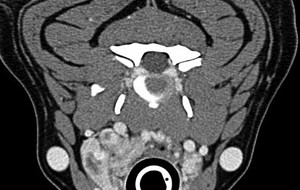
This month's CT/MRI Society case is from Shannon Holmes at the University of Georgia. Just a reminder that these cases are available to view for 1 month only. If you'd like to have access to all of them, become a member of the ACVR CT/MRI Society. Enjoy! Access full case information and all … [Read more...]
- 1
- 2
- 3
- 4
- Next Page »