• Home
  • About
  • Articles
  • Books
  • Contact

Veterinary Radiology

Teaching and learning about veterinary diagnostic imaging.

  • Modality
    • Radiographs
    • Ultrasound
    • CT/MRI
  • Region
    • Thorax
    • Abdomen
    • Musculoskeletal
    • Neurologic
  • Species
    • Canine
    • Feline
    • Equine
    • Exotic

2 year old Pit Bull terrier

February 25, 2016 By Allison Zwingenberger Leave a Comment

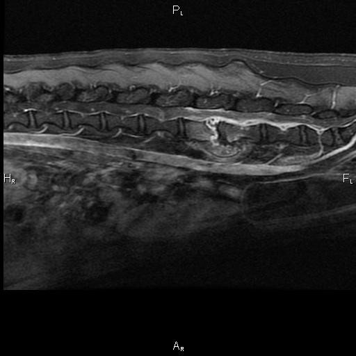
Today’s case is a 2-year-old female neutered Pit bull terrier. Fell off bed 10 days previously, acute paraparesis. What are your findings?

L LAT Spine
L LAT Spine
L LAT Spine
L LAT Spine
L LAT Spine
L LAT Spine
L LAT Spine
L LAT Spine
L LAT Spine
VD Spine
VD Spine
VD Spine
VD Spine

Show findings...

Findings

There is a fracture of the caudal end plate of the L4 vetebral body with adjacent sclerosis and lysis. There is ventral spondylosis deformans at this site. Mineral opacity material is present in the intervertebral foramen. The remainder of the vertebral bodies appear normal. On flexed and extended views, there is no evidence of instability.

Show diagnosis…

Diagnosis

Fracture and osteomyelitis. It is unclear which pathology is primary, and a migrating foreign body could also be involved.

Show discussion…

Discussion

An MRI was performed confirming the osteolysis and inflammatory changes in the vertebral body. Contrast enhancing tissue consistent with empyema was visible involving the meninges, as well as inflammation of the musculature and soft tissues.

MRI_lat_c

Filed Under: Canine, Case of the Day, CT/MRI, Musculoskeletal, Radiographs

Leave a Reply Cancel reply

You must be logged in to post a comment.

  • Bloglovin
  • Facebook
  • RSS
  • Twitter
AtlasCover787x1024 amazon-availablenow

Veterinary Radiology News

Sign up for alerts about new cases and newsletters.

We will respect your privacy.

vetHP Osirix Plugin

Save time, read more cases! VetHP automatically changes your CT and MR images to veterinary orientation.
Improve your workflow today!
vethp

Archives

Recent Comments

  • filipp: Good case, I missed the linear foreign body in the trachea
  • filipp: Great case
  • logan: How can we be positive about that opacity on the 6th ribs being nothing more that a summation artifact?
  • edevis: Thank you for the interesting case again. A non “radiologic” comment: vizsla is spelled with the...
  • Anna N: Hi, I was just wandering what this ‘ linear mineral density separated from the joint surface’...

Tags

adenocarcinoma bronchiectasis carcinoma cardiomyopathy cardiovascular coccidioidomycosis discospondylitis elbow dysplasia eosinophilic bronchopneumopathy erosive polyarthritis esophageal foreign body FIP foreign body fracture gastrointestinal GDV genitourinary heart failure heartworm hemangiosarcoma hiatal hernia hypertrophic osteodystrophy intestinal foreign body laryngeal paralysis linear foreign body lipoma lymphoma megaesophagus metastatic disease osteochondrosis osteomyelitis osteosarcoma patent ductus arteriosus PDA pericardial effusion pneumonia pneumothorax polyarthritis PPDH pulmonary pulmonary abscess pulmonary adenocarcinoma sarcoma tracheal collapse vascular ring anomaly

Copyright © 2018 · Metro Pro Theme On Genesis Framework · WordPress · Log in