This is a great case of breed-related disease. Post your interpretations in the comments section.
Access all images and case information.
Case originally posted on June 12, 2008
Teaching and learning about veterinary diagnostic imaging.
This is a great case of breed-related disease. Post your interpretations in the comments section.
Access all images and case information.
Case originally posted on June 12, 2008
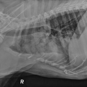
There are some sreas of soft tissue opacities in the right cranial lung lobe and mediastinum, and some similar ill defined soft tissue opacities accompined by alveolar infiltrates in the left caudal lung field. With the signalment malignant hystiocytosis is very likely. Other neoplastic processes should also be considered. An aspirate is recommended.
Posted on behalf of Dennis:
When reviewing these rads I see
1. See some arthritic change in the shoulder
2. Is this a lytic bone on rt lateral 4th dorsal process… not seeing on the left and don’t think an issue
3. Multiple nodular densities in cranial lung lubes, is in caudal somewhat also
* Is rt cranial mass .. off mediastinum or rt cranial lobe .. think mediastinum
* Some alveolar mass in left cranial lobes also
* Mixed interstitial alveolar pattern more caudally
4. Pleural fluid line
R/O Hemorr Abcess Neoplasia Granuloma in the lung ..// mediastinum think Neoplasia Granuloma
Slightly rotated VD view. Slight terminal trachea ventral deviation on the rt latelar view could manifest carina l/n enlargement. Soft tissue mass in the rt cr thorax without pleural sign and silhouette sign with the heart. The mass is cavitated (air), does not deviates trachea or shifts the mediastinum. I think is lung mass. Rounded soft tissue masses in the caudal aspect of the lt cr lobe and in the lt caudal lobe. Semi-oval mass in the lt caudal thorax projecting from the lt diaphragmatic crus (hernia??). Rounded opaque mass in the region of the lt liver next to the stomach. Bronchiectasis of the lt cranial lung lobe bronchus. Bronchointerstitial pattern in the rt medial and caudal lobes and lt cranial lobe. Pleural fissure lines in the rt caudal and lt cr thorax. Some osteophytes in the rt shoulder.
Considering the history R/O: Neoplasia, Abscess, Hystiocytosis, Myasthenia gravis
Good job! Everyone agrees that there are multiple lung masses. Before you look at the answers (click on the link to the case above), try to determine which lung lobes are involved. If the findings could be due to a number of diseases, a good strategy is to stay broad and list categories such as abscess, granuloma, neoplasia etc. If you are more sure that it’s neoplasia, try to create an ordered list of possible diagnoses. Your differentials depend on your level of certainty for a particular finding or case.
Okay, I think we have cranio ventral increase opacity in the torax, dorsal diplasment of trochea, multiole masses in the lung, I think intersticial pathern in dorsal caudal lung, I think it could be right heart hemangiosarcoma too.
Hypoventilation, upper or lower airway obstruction, pleural filling disorders, pulmonary parenchymal.