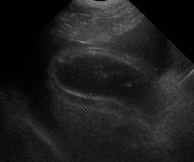
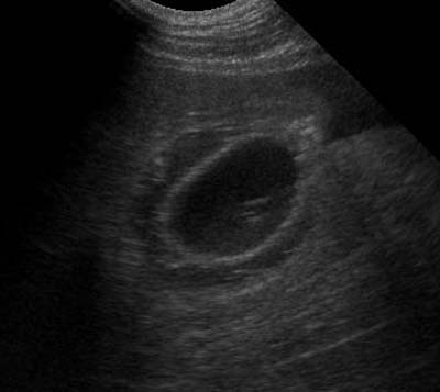
Edema of the gall bladder wall looks like a hypoechoic layer between two hyperechcoic surfaces. It can be confused with a small amount of peritoneal effusion so look carefully at the neck and body. In the images here, you can see the anechoic peritoneal effusion surrounding the thickened gall bladder wall.
Although gall bladder wall edema can be seen with inflammatory hepatic and biliary disease, it’s also associated with other conditions including hypoalbuminemia and sepsis. These sagittal (image 1) and transverse (image 2) images of the gall bladder show edema induced by sepsis. In my experience, the edema can resolve quickly if the systemic illness improves.
Spaulding Kathy A. Gallbladder wall thickness – Ultasound corner. Veterinary Radiology & Ultrasound 1993;34:270-272.
Recent Comments