• Home
  • About
  • Articles
  • Books
  • Contact

Veterinary Radiology

Teaching and learning about veterinary diagnostic imaging.

  • Modality
    • Radiographs
    • Ultrasound
    • CT/MRI
  • Region
    • Thorax
    • Abdomen
    • Musculoskeletal
    • Neurologic
  • Species
    • Canine
    • Feline
    • Equine
    • Exotic

4 month old Jack Russell Terrier

April 5, 2018 By Allison Zwingenberger 1 Comment

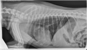
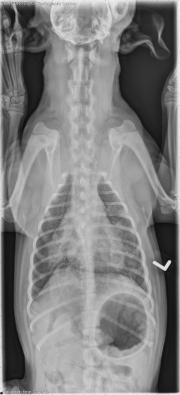
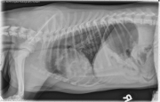
Today’s case is a 4-month-old Jack Russell Terrier with wheezing and coughing. Three puppies were placed in a stall on cedar wood shavings to keep them warm. When the owner returned home that evening, she noticed that this puppy was wheezing and coughing. A chewed collar was found in the shavings. Take a look and post your comments!
cod180405-01
cod180405-02
cod180405-03
cod180405-04
Show findings...

Findings

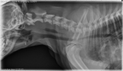
On radiographs of the thorax, there is an alveolar pulmonary pattern in the left cranial, and right middle lung lobes, with patchy increased opacity in the remaining lobes. The trachea is narrowed at the thoracic inlet, with an apparent linear intraluminal opacity. There is dilation of the pharynx with air, and there is gas within the esophagus and gastrointestinal tract.

Show differential diagnosis…

Differential Diagnosis

The tracheal narrowing and intraluminal opacity may indicate edema, mucus, or foreign material in this region. There is secondary upper airway obstruction as indicated by the dilated pharynx and aerophagia. The alveolar pattern is due to bronchopneumonia.

Show diagnosis…

Diagnosis

Endoscopy was performed, and a 7 inch plant foreign body was removed from the main stem bronchus of the left caudal lung lobe. The trachea was inflamed, and the bronchoalveolar lavage showed moderate to marked, septic, suppurative inflammation and hemorrhage, with small numbers of Pasteurella and Mycoplasma cultured. The patient improved on antibiotic therapy and supportive care and was discharged.

Filed Under: Canine, Case of the Day, Radiographs, Thorax

Comments

  1. filipp says

    April 5, 2018 at 4:23 am

    Good case, I missed the linear foreign body in the trachea

    Log in to Reply

Leave a Reply Cancel reply

You must be logged in to post a comment.

  • Bloglovin
  • Facebook
  • RSS
  • Twitter
AtlasCover787x1024 amazon-availablenow

Veterinary Radiology News

Sign up for alerts about new cases and newsletters.

We will respect your privacy.

vetHP Osirix Plugin

Save time, read more cases! VetHP automatically changes your CT and MR images to veterinary orientation.
Improve your workflow today!
vethp

Archives

Recent Comments

  • filipp: Good case, I missed the linear foreign body in the trachea
  • filipp: Great case
  • logan: How can we be positive about that opacity on the 6th ribs being nothing more that a summation artifact?
  • edevis: Thank you for the interesting case again. A non “radiologic” comment: vizsla is spelled with the...
  • Anna N: Hi, I was just wandering what this ‘ linear mineral density separated from the joint surface’...

Tags

adenocarcinoma bronchiectasis carcinoma cardiomyopathy cardiovascular coccidioidomycosis discospondylitis elbow dysplasia eosinophilic bronchopneumopathy erosive polyarthritis esophageal foreign body FIP foreign body fracture gastrointestinal GDV genitourinary heart failure heartworm hemangiosarcoma hiatal hernia hypertrophic osteodystrophy intestinal foreign body laryngeal paralysis linear foreign body lipoma lymphoma megaesophagus metastatic disease osteochondrosis osteomyelitis osteosarcoma patent ductus arteriosus PDA pericardial effusion pneumonia pneumothorax polyarthritis PPDH pulmonary pulmonary abscess pulmonary adenocarcinoma sarcoma tracheal collapse vascular ring anomaly

Copyright © 2018 · Metro Pro Theme On Genesis Framework · WordPress · Log in