The pet food recall has been the primary topic in veterinary related news for several months. The substance identified in the renal tubules of affected animals' kidneys is melamine, used in fertilizers and plastics. The doses found in contaminated feeds are low, and it's still unclear whether this … [Read more...]
Do advertisements work? fMRI investigates.
We all think of MR as a diagnostic tool to let us look inside the brain for signs of disease. It gives fantastic anatomical detail and contrast between tissues. Functional MR, or fMRI, is becoming a popular tool in investigating how the brain works. It measures changes in blood flow in different … [Read more...]
The Pet Owner’s Guide to Veterinary Radiology
It's interesting to see what people are looking for when they land on the Veterinary Radiology home page. In this post, I'll address some of the more common questions that people want answered about our specialty. What is a veterinary radiologist? A veterinary radiologist is a veterinarian who has … [Read more...]
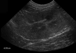
Splenic nodules
Splenic nodules or masses are extremely common findings when ultrasounding the canine abdomen. Nodules are small, circular abnormalities within the spleen that might be hyperechoic (brighter) or hypoechoic (darker) than the surrounding, normal spleen. Nodules are small (less than 4 cm diameter) and … [Read more...]
Melamine identified as potential toxin in pet food recall
Melamine has been identified in the recalled pet food, as well as in the urine and kidneys of an affected animal. The chemical, used in plastics, has been found in the contaminated gluten used in wet pet foods. However, there may have been contamination of dry foods as well. You can read more about … [Read more...]
Recent Comments