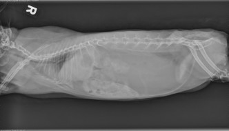
Archive for the 'Known Case Conference' Category

This week’s collection of cases was a mixture - from GI to large animal radiographs to thorax. As usual, the residents did a great job of describing the findings, coming up with a radiologic diagnosis, and a list of differential diagnoses. These files are digital so make sure to look at the images as well.

Case 1

10 year old Bull mastiff with 3 weeks of intermittent vomiting. There is a severely dilated bowel loop in the dorsal right abdomen that contains striated foreign material. There is a tortuous, gas dilated loop cranial to this loop. The stomach apppears small. The peritoneal detail is poor. Differentials include chronic partial obstruction secondary to a stricture or mass, or a textile foreign body. Poor detail may indicate peritonitis from inflammation or bowel permeability. There is no free gas. Diagnosis: Schirrous adenocarcinoma with secondary chronic partial obstruction. Tip: Older dogs are less likely to ingest foreign bodies, so be on the lookout for causes of obstruction such as strictures or masses in the GI tract.

Case 2

Equine shoulder radiographs. 27 year old Arab gelding found non-weight bearing. The distal lateral radiograph of the shoulder did not include the glenohumeral joint. However, there is a possible bone fragment at the caudal aspect of the proximal humerus which is not visible on the more proximal views. Differentials include an artifact or a fracture fragment. Request additional radiographs more proximally with increased exposure. These, taken at a cranial 30 degrees medial to caudolateral oblique angle, demonstrated a fracture of the humeral tubercle with moderate caudal and distal displacement. Horses have a medial, central and lateral tubercle of the humerus. Diagnosis: Slab fracture of the humeral head parasagittal with medial tubercle fracture.

Case 3

5 year Arabian gelding. Large swelling with draining wound medial to left stifle. The radiographs are centered on the soft tissue swelling rather than the stifle. There is some motion artifact because the horse was painful. There is a metallic marker delineating the location of the wound. Differentials include hematoma or abscess. There is a linear lucent area in the soft tissues. Diagnosis: 6 inch wooden foreign body..

Case 4

1 year Yorkshire Terrier with polycythemia (77%). On thoracic radiographs, there is marked right sided cardiac enlargement. The aortic arch is enlarged on the d/v projection. One of the caudal lobar pulmonary arteries is blunted which may indicate pulmonary thromboembolism. The diagnosis is a congenital cardiac anomaly. Differentials include a right to left shunt including PDA, VSD, tetralogy of Fallot. Next steps could include non-selective angiography or echocardiography with bubble study, or nuclear scintigraphy. Using echocardiography, bubbles will bypass the lungs and appear in the abdominal aorta.

Case 5

2 month old St Bernard found wet and cold in the house (no images available). The patient presented dyspneic and was placed on the ventilator. Thoracic radiographs showed an alveolar pulmonary pattern in the caudodorsal lungs. A catheter is present in the cranial vena cava and there is an endotracheal tube in the trachea. There are also electrical leads on the chest wall. The most likely differential diagnosis is non-cardiogenic pulmonary edema secondary to asphyxiation, near drowning or electrical shock. Embolic pneumonia is less likely. Diagnosis: Non-cardiogenic pulmonary edema from falling in swimming pool.

This week at KCC we had some interesting and unusual cases. They were all on film, so I’m afraid there aren’t any radiographs to look at. Read the description and decide what you would do before reading the answer. In these types of cases, you aren’t expected to get “the answer” but to provide a logical analysis and appropriate differentials.

Mature domstic short hair cat with 3 week history of respiratory distress

Thoracic radiographs showed a flattened diaphragm and hyperlucent lungs. On the d/v projection, the ribs were nearly perpendicular to the spine and the diaphragm had scalloped edges. The esophagus was markedly dilated with air, as was the stomach and small intestine. These findings of hyperinflation and aerophagia suggested an upper airway obstruction so neck radiographs were requested. In the larynx, there was a soft tissue opacity mass that was responsible for the upper airway obstruction. Differential diagnoses included neoplasia (lymphoma, squamous cell carcinoma), granulomatous disease, and edema. Diagnosis: lymphoma.

9 month old domestic long hair cat with a heart murmur

The heart was markedly enlarged on both the lateral and d/v projections. The shape of the cardiac silhouette resembled a peanut with a waist between the enlarged atria and ventricles. The heart occupied more than 50% of the thorax on the d/v projection. The pulmonary arteries and veins were enlarged. On the edge of the radiograph, the fundus of the stomach was visible on the right side. Differential diagnoses included congenital heart disease such as vsd and abnormal valves, as well as PDA or VSD causing a left to right shunt. Secondary diagnosis: Situs inversus. Echocardiography revealed a single atrium and single ventricle with a VSD. Note: This type of case is not well diagnosed on radiographs beyond severe, congenital heart disease. Complex cardiac anomalies can be associated with situs inversus.

23 year old Morgan gelding with anorexia and weight loss

Thoracic ultrasound: There was marked echogenic pleural effusion with visible strands of the pericardial-diaphragmatic ligament. In the cranial thorax, there was a lobular mass with relatively uniform echogenicity. Differential diagnoses for the mass were neoplasia, most likely lymphoma, hemangiosarcoma, melanoma, or other neoplastic disease. Pleural fluid was non-diagnostic. A tissue sample was obtained by thoracoscopy. Diagnosis: anaplastic carcinoma.

This week’s KCC was about practicing interpreting an ultrasound video. It’s part of the oral exam, and it can be very disconcerting to watch someone else “drive”. There’s always something you want to see that they keep moving past! But it’s good practice to do the interpretations to orient yourself, describe what you see, and put it together with the history to make your interpretation.

Case 1

13 year old Siamese cat with lethargy. This scan started at the liver, which was enlarged and hypoechoic. The gall bladder was moderately distended, as was the common bile duct. The CBD was tortuous and looped back on itself before joining the duodenal papilla. There was no obvious mass at the papilla. The pancreas was mildly enlarged and hypoechoic but did not appear inflamed. The pancreatic duct was also enlarged. The diagnosis was chronic partial obstruction of the common bile duct, and differentials included inflammatory disease, neoplasia or calculus. The liver and pancreatic changes suggested chronic inflammation as is common in this group of diseases in cats. One year later, the changes looked similar, so inflammatory disease was the most likely diagnosis.

Case 2

11 year old Golden Retriever who had a red eye for two weeks and now has a painful eye. The ultrasound scanned through the globe. The anterior chamber and lens were identified. The vitreous body contained a hyperechoic, round mass that appeared to have strands connecting it to the iris and also touched the choroid at the back of the globe. The retina was not visible and the vitreous was clear. The most likely differential was neoplasia, since the vitreous was anechoic and showed no evidence of inflammation. Possible origins of the mass were the iris (melanoma) and the choroid. Diagnosis: melanoma originating from the iris.

Case 3

7 year old bull terrier with a mass on the neck. Orientation was difficult in this case. The scan appeared to be from lateral, with the mandibular salivary gland visible with a spleen-like echotexture in the cranial portion of the image. Caudal to the gland, there was a thin-walled, fluid-filled structure. The fluid was echogenic and the cystic area had complex septations. There were several small calculi in the far field that produced acoustic shadowing. There was a duct leading from the salivary gland into the cyst and it contained a calculus. Diagnosis: sialocele with sialoliths.